

 PDF
PDF ePub
ePub Citation
 Print
Print


Abstract
Purpose
To report a case of a young male patient with retinitis pigmentosa (RP) accompanied by vitritis and neo-vascularization of the optic disk in both eyes who underwent unilateral vitrectomy for the treatment of vitreous hemorrhage in the right eye.
Case summary
An 8-year-old boy visited our clinic with a complaint of night blindness. Both eyes showed inflammatory cells in the anterior vitreous and neovascularization of the optic disk confirmed by fluorescein angiography. Extensive vitreous hemorrhage developed in his right eye and he underwent unilateral vitrectomy. His final visual acuity was 0.6 in both eyes.
Go to : 
References
1. Wong P, Borst DE, Farber D. . Increased TRPM-2/clusterin mRNA levels during the time of retinal degeneration in mouse models of retinitis pigmentosa. Biochem Cell Biol. 1994; 72:(9-10). 439–46.

2. Cotran PR, Bruns GA, Berson EL, Dryja TP. Genetic analysis of patients with retinitis pigmentosa using a cloned cDNA probe for the human gamma subunit of cyclic GMP phosphodiesterase. Exp Eye Res. 1991; 53:557–64.
3. Uliss AE, Gregor ZJ, Bird AC. Retinitis pigmentosa and retinal neovascularization. Ophthalmology. 1986; 93:1599–603.
4. Newsome DA, Michels RG. Detection of lymphocytes in the vitre-ous gel of patients with retinitis pigmentosa. Am J Ophthalmol. 1988; 105:596–602.
5. Yoshida N, Ikeda Y, Notomi S. . Clinical evidence of sustained chronic inflammatory reaction in retinitis pigmentosa. Ophthalmology. 2013; 120:100–5.
6. Nao-i N, Fukiyama J, Sawada A. Retinitis pigmentosa with re-current vitreous hemorrhage. Acta Ophthalmol Scand. 1996; 74:509–12.
7. Bressler NM, Graqoudas ES. Neovascularization of the optic disk associated with atypical retinitis pigmentosa. Am J Ophthalmol. 1985; 100:431–3.
8. Henkind P. Ocular neovascularization. The Krill memorial lecture. Am J Ophthalmol. 1978; 85:287–301.
Go to :
 | Figure 1.(A, B) Fundus photographs show diffuse retinal degeneration with sparing of the macula in both eyes. (C, D) Electroretinograms showing nearly reduced ‘a’ and ‘b’ waves in the both eyes. |
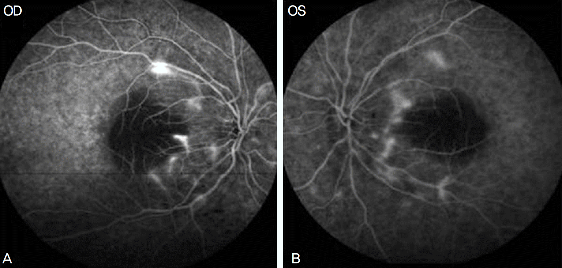 | Figure 2.(A, B) Fluorescein angiogram of the both eyes show dye leakage from new vessels around the optic disc margin. |

 XML Download