

 PDF
PDF ePub
ePub Citation
 Print
Print


Abstract
Purpose
To report a case of orbital apex syndrome induced by penetrating orbital injury by a wire with the recovery proc-ess and clinical outcomes.
Case summary
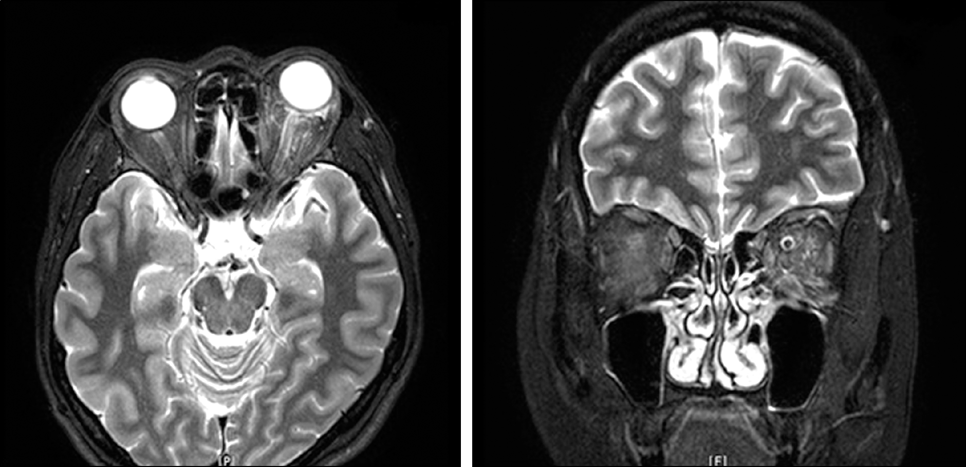
A 40-year-old female visited our clinic after a penetrating orbital injury through the left inferomedial con-junctiva by a wire. The best corrected visual acuity of the left eye was 0.6, and ptosis and total ophthalmoplegia were observed. The patient showed a dilated pupil, swelling of the optic disc on fundus exam, and an inferior field defect on the automated perimetry. The computed tomography image revealed mild retrobulbar hemorrhage, but there was no orbital bony fracture. Enhancement of the optic nerve sheath was observed on the magnetic resonance image. The patient was admitted and received systemic antibiotics and steroid treatment. After 1 month, visual acuity, ptosis, and limitation in ad-duction were partly improved. After 3 months, depression and adduction were improved and the pupil size was normalized. However, further improvement was not observed after the one-year follow-up.
References
1. Zachariades N, Vairaktaris E, Papavassiliou D. . Orbital apex syndrome. Int J Oral Maxillofac Surg. 1987; 16:352–4.

2. Bray WH, Giangiacomo J, Ide CH. Orbital apex syndrome. Surv Ophthalmol. 1987; 32:136–40.
3. Brent BD, May DR. Orbital apex syndrome after penetrating orbi-tal trauma. Ann Ophthalmol. 1990; 22:267–8.
4. Cho EY, Choi YK, Choi WC. A case of nasal endoscopic treatment for paranasal mucocele. J Korean Ophthalmol Soc. 2004; 45:1386–91.
5. Baik SH, Yeom DJ, Kang YK. . Orbital apex syndrome with nasal type natural killer(NK)/T-cell lymphoma of sphenoid and ethmoid sinus. J Korean Ophthalmol Soc. 2010; 51:286–91.
6. Kim TH, Lee EC, Seo SW, Yoo JM. Two case of orbital apex syndrome. J Korean Ophthalmol Soc. 2002; 43:927–33.
7. Steinsapir KD, Goldberg RA. Traumatic optic neuropathy: Perspective traumatic optic neuropathy: An evolving understanding. Am J Ophthalmol. 2011; 151:928–33.e2.
8. Keane JR. Cavernous sinus syndrome. Analysis of 151 cases. Arch Neurol. 1996; 53:967–71.
9. Lenzi GL, Fieschi C. Superior orbital fissure syndrome. Review of 130 cases. Eur Neurol. 1977; 16:23–30.
10. Garland SD, Maloney PL, Doku HC. Carotid-cavernous sinus fis-tula after trauma to the head. J Oral Surg. 1977; 35:832–5.
11. Lew H, Lee SY, Jang JW. . The effect of high dose cortico-steroid therapy on the optic nerve head blood flow in experimental traumatic optic neuropathy. J Korean Ophthalmol Soc. 1998; 39:1771–8.
12. Levin LA, Beck RW, Joseph MP. . The treatment of traumatic optic neuropathy: the International Optic Nerve Trauma Study. Ophthalmology. 1999; 106:1268–77.
13. Braken MB, Shepard MJ, Holford TR. . Administration of methylprednisolone for 24 or 48 hours or tirilazad mesylate for 48 hours in the treatment of acute spinal cord injury. Results of the Third National Acute Spinal Cord Injury Randomized Controlled Trial. National Acute Spinal Cord Injury Study. JAMA. 1997; 277:1597–604.
14. Roberts I, Yates D, Sandercock P. . Effect of intravenous corti-costeroids on death within 14 days in 10008 adults with clinically significant head injury (MRC CRASH trial): randomised place-bo-controlled trial. Lancet. 2004; 364:1321–8.
15. Spoor TC, Hartel WC, Lensink DB, Wilkinson MJ. Treatment of traumatic optic neuropathy with corticosteroids. Am J Ophthalmol. 1990; 110:665–9.
16. Bracken MB, Shepard MJ, Collins WF. . A randomized, con-trolled trial of methylprednisolone or naloxone in the treatment of acute spinal-cord injury. Results of the Second National Acute Spinal Cord Injury Study. N Engl J Med. 1990; 322:1405–11.
17. Medeiros FA, Moura FC, Vessani RM, Susanna R Jr. Axonal loss after traumatic optic neuropathy documented by opticalcoherence tomography. Am J Ophthalmol. 2003; 135:406–8.
Figure 1.
Fields of gazes on initial presentation show limitation of the extraocular muscle movement in all directions. Complete pto-sis of the left upper eyelid is also noted.

Figure 2.
(A) Optic disc photograph, OCT, and visual field test findings on initial presentation. Optic disc photograph (upper left) demonstrates whitish elevation at the superior disc margin. OCT (upper middle) findings show increase thickness of the superior RNFL which correlated with the optic disc photographs. Automated perimetry (upper right) reveals inferior altitudinal visual field defects on the gray-scale. (B) One month after injury, optic disc photograph shows no significant change. However, OCT shows the thickness of superior RNFL reduced to the normal range, and the visual field defects also improved accordingly. OCT = optical co-herent tomography; RNFL = retinal nerve fiber layer.

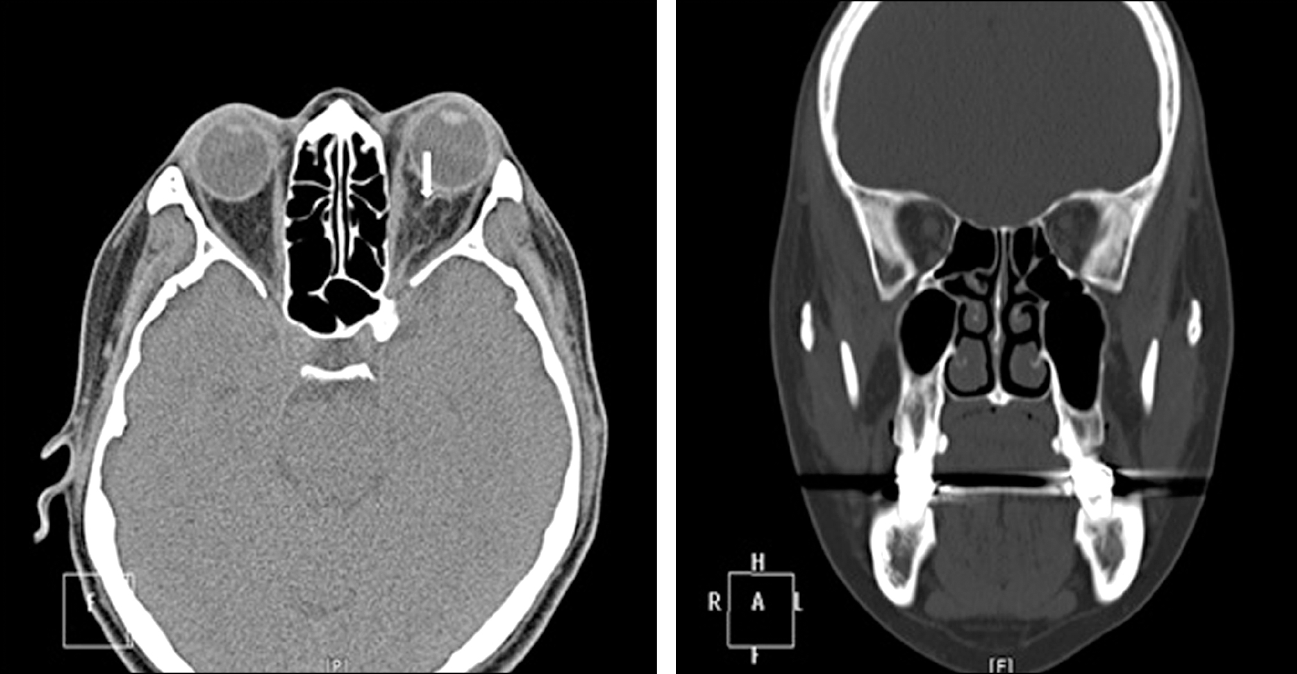
Figure 3.
Axial (left) and coronal (right) CT scan on initial presentation. CT demonstrates an increased reticular pattern with-in the right intraconal fat and around the postlaminar optic nerve suggesting mild retrobulbar hemorrhage (arrow). There is no orbital bony fracture.

 XML Download