Abstract
Purpose
To evaluate the incidence and course of widening of palpebral fissure after unilateral lateral rectus muscle recession.
Methods
The palpebral fissure width (PFW) was measured in 20 patients with intermittent exotropia before unilateral rec-tus muscle recession and 1 week, 1 month and 6 months after the surgery.
Results
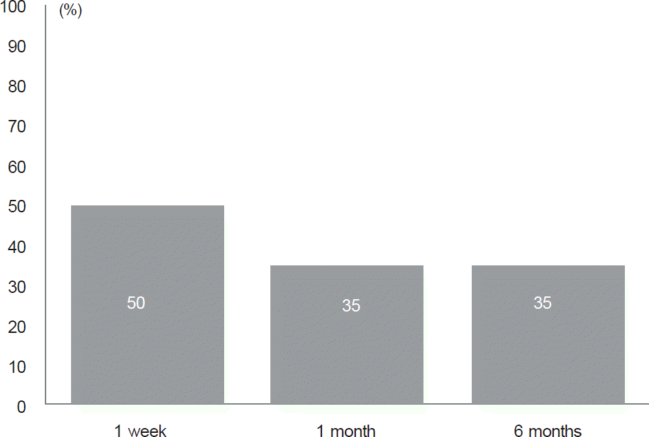
The amount of recession was from 7.5 to 9.0 (mean 8.37 ± 0.51) mm. More than 0.6 mm of change in PFW after surgery was defined as the significant change. The significant change was observed in 10 patients (50%) after 1 week, 7 patients (35%) after 1 month and 7 patients (35%) after 6 months after the surgery. The amount of recession was sig-nificantly greater in the group with significant change (8.60 ± 0.39 mm) than the group without significant change (8.15 ± 0.53 mm) (p < 0.05).
Conclusions
Thirty five percent of the patients showed palpebral fissure widening lasts at least 6 months after unilateral lateral rectus muscle recession. We think it is necessary to notice patients about the possible change in palpebral fissure width before strabismus surgery. And we believe that more cosmetically satisfactory outcome would be resulted if surgeons consider eyelid condition when they are planning strabismus surgery.
Go to : 
References
1. Lueder GT, Scott WE, Kutschke PJ, Keech RV. Long-term results of adjustable suture surgery for strabismus secondary to thyroid ophthalmopathy. Ophthalmology. 1992; 99:993–7.

2. Pacheco EM, Guyton DL, Repka MX. Changes in eyelid position accompanying vertical rectus muscle surgery and prevention of lower lid retraction with adjustable surgery. J Pediatr Ophthalmol Strabismus. 1992; 29:265–72.

3. Lam BL, Lam S, Walls RC. Prevalence of palpebral fissure asym-metry in white persons. Am J Ophthalmol. 1995; 120:518–22.

4. Kwak CY, Yu YS. Analysis of lid contours in children. J Korean Ophthalmol Soc. 1991; 32:520–6.
5. McCracken MS, del Prado JD, Granet DB. . Combined eyelid and strabismus surgery: examining conventional surgical wisdom. J Pediatr Ophthalmol Strabismus. 2008; 45:220–4.

6. Lagrèze WA, Gerling J, Staubach F. Changes of the lid fissure after surgery on horizontal extraocular muscle. Am J Ophthalmol. 2005; 140:1145–6.
7. Santos de Souza Lima LC, Velarde LG, Vianna RN. . The ef-fect of horizontal strabismus surgery on the vertical palpebral fis-sure width. J AAPOS. 2011; 15:473–5.

8. Chen SI, Knox PC, Hiscott P, Marsh IB. Detection of the slipped extraocular muscle after strabismus surgery. Ophthalmology. 2005; 112:686–93.

Go to : 
 | Figure 1.Measurement of the Palpebral fissure width. We measured the distance between upper and lower eyelid margin by placing the image of reference ruler on point of corneal re-flex after cut off it on the same photograph. |
 | Figure 2.Incidence rate of the significant change of palpebral fissure width at each follow up point. |
Table 1.
Demographics of the subjects
Table 2.
The differences between both eyes and amount of change in palpebral fissure width (PFW) and mean width of operated eye (mm)
| Subjects | Baseline | 1 week | 1 month | 6 months |
|---|---|---|---|---|
| 1 | 0.4 | 1.6 (1.2)* | 1.6 (1.2)* | 1.6 (1.2)* |
| 2 | 0 | 0.6 (0.6)* | 0.6 (0.6)* | 0.6 (0.6)* |
| 3 | 0 | 1 (1)* | 0.8 (0.8)* | 0.8 (0.8)* |
| 4 | 0 | 0.6 (0.6)* | 0 (0) | 0 (0) |
| 5 | 0 | 0.6 (0.6)* | 0 (0) | 0 (0) |
| 6 | 0 | 1 (1)* | 1 (1)* | 0.6 (0.6)* |
| 7 | 0 | 0.6 (0.6)* | 0.8 (0.8)* | 0.8 (0.8)* |
| 8 | 0 | 0.6 (0.6)* | 1 (1)* | 1 (1)* |
| 9 | 0 | 0.6 (0.6)* | 1 (1)* | 1 (1)* |
| 10 | 0 | 0 (0) | 0 (0) | 0 (0) |
| 11 | 1 | 1 (0) | 1 (0) | 1 (0) |
| 12 | 0.4 | 0.6 (0.2) | 0.6 (0.2) | 0.4 (0) |
| 13 | 0 | 0 (0) | 0 (0) | 0 (0) |
| 14 | 0 | 0 (0) | 0 (0) | 0 (0) |
| 15 | 0 | 0 (0) | 0 (0) | 0 (0) |
| 16 | 0.4 | 0.4 (0) | 0.4 (0) | 0.4 (0) |
| 17 | 0.4 | 0.4 (0) | 0.4 (0) | 0.4 (0) |
| 18 | 0.2 | 0.8 (0.6)* | 0.4 (0.2) | 0.4 (0.2) |
| 19 | 0.4 | 0.4 (0) | 0.4 (0) | 0.4 (0) |
| 20 | 0.2 | 0.2 (0) | 0.2 (0) | 0.2 (0) |
| Mean PFW | 7.76 ± 1.04 | 8.05 ± 1.01 | 8.13 ± 0.99 | 8.04 ± 0.80 |
Table 3.
Comparison of parameters between the group with the significant change and the group without significant change
| Significant change | No significant change | p-value* | |
|---|---|---|---|
| Age (years) | 7.0 ± 1.56 | 8.5 ± 3.77 | 0.529 |
| Preoperative deviation (prism diopter) | 21.2 ± 3.61 | 17.0 ± 2.53 | 0.015 |
| Amount of LR recession (mm) | 8.60 ± 0.39 | 8.15 ± 0.53 | 0.048 |


 PDF
PDF ePub
ePub Citation
 Print
Print


 XML Download