

 PDF
PDF ePub
ePub Citation
 Print
Print


Abstract
Purpose
To evaluate clinical patterns according to the occlusion site in patients with branch retinal vein occlusion.
Methods
Ninety-one branch retinal vein occlusion patients were divided into 4 groups according to the occlusion site based on the description by Duke-Elder and Wybar: Papillary retinal vein occlusion group (group A), main retinal vein oc-clusion group (group B), minor retinal vein occlusion group (group C), and retinal venule occlusion group (group D). The following factors were analyzed retrospectively: baseline/final visual acuity, visual improvement, macular thickness, and macular circulatory states.
Results
The macular thickness (μ m) was 575.33 ± 178.44 in group A, 511.92 ± 218.02 in group B, 397.21 ± 144.51 in group C, and 336.68 ± 120.55 in group D. The retinal ischemic area (DD) was 22.00 ± 13.28 in group A, 18.26 ± 10.12 in group B, 12.52 ± 10.52 in group C, and 12.36 ± 11.92 in group D, which was found to be significantly greater in the group with the higher branch occlusion site (p < 0.05). However, visual acuity, macular circulatory states and other clinical char-acteristics were not significantly different.
References
1. Orth DH, Patz A. Retinal branch vein occlusion. Surv Ophthalmol. 1978; 22:357–76.

2. Roseman RL, Olk RJ. Krypton red laser photocoagulation for branch retinal vein occlusion. Ophthalmology. 1987; 94:1120–5.
3. Finkelstein D. Retinal branch vein occlusion. In: Ryan SJ, eds. Retina. 2nd ed.St. Louis: Mosby;1994; 1387–92.
4. Wilson DJ, Finkelstein D, Quigley HA, Geen WR. Macular grid photocoagulation. An experimental study on the primate retina. Arch Ophthalmol. 1988; 106:100–5.
5. Min WK, Hong ST, Park YG, Park KS. The natural course and visual prognosis of branch retinal vein occlusion. J Korean Ophthalmol Soc. 1994; 35:664–72.
6. Shilling JS, Jones CA. Retinal branch vein occlusion: A Study of Argon laser photocoagulation in the treatment of macular oedema. Br J Ophthalmol. 1984; 68:196–8.
7. Son MH, Yoon IH. Macular ischemia in temporal branch retinal vein occlusion fluorescein angiographic classification and visual prognosis. J Korean Ophthalmol Soc. 1996; 37:608–13.
8. Clemett RS, Kohner EM, Halmilton AM. The visual prognosis in retinal branch vein occlusion. Trans Ophthalmol Soc U K. 1973; 93:523–35.
9. Beaumont PE, Kang HK. Clinical characteristics of retinal venous occlusions occurring at different sites. Br J Ophthalmol. 2002; 86:572–80.
10. Chung JH, Choi GJ, Na KS. The macular circulation state on BRVO according to occlusion site. J Korean Ophthalmol Soc. 2000; 41:1556–62.
11. Kim HC, Kim PS, Kim HK. The macular circulatory change in branch retinal vein occlusion. J Korean Ophthalmol Soc. 1999; 40:1911–7.
12. Kang SJ, Chin HS, Moon YS. Visual prognosis of macular edema associated with macular ischemia in branch retinal vein occlusion. J Korean Ophthalmol Soc. 2002; 43:1621–8.
13. Duke-Elder S, Wybar KC. The anatomy of the visual system. In: Duke-Elder, eds. System of Ophthalmology. St Louis: Mosby;v. 2:1961–390.
14. Hill DW, Griffiths JD. The prognosis in retinal vein thrombosis. Trans Ophthalmol Soc U K. 1970; 90:309–22.
15. Finkelstein D. Ischemic macular edema. Recognition and favor-able natural history in branch vein occlusion. Arch Ophthalmol. 1992; 110:1427–34.
16. Gutman FA, Zegarra H. The natural course of temporal retinal branch vein occlusion. Trans Am Acad Ophthalmol Otolaryngol. 1974; 78:178–92.
17. Gutman FA. Macular edema in branch retinal vein occlusion: prog-nosis and management. Trans Sect Ophthalmol Am Acad Ophthalmol Otolaryngol. 1977; 83:(3 Pt 1). 488–95.
18. The Branch Vein Occlusion Study Group . Argon laser photo-coagulation for macular edema in branch vein occlusion. Am J Ophthalmol. 1984; 98:271–82.
19. Jalkh AE, Trempe CL. Macular edema in branch retinal vein occlusion: types and treatment. Ophthalmic Surg. 1989; 20:26–32.
20. Rehák J. [Retinal vein occlusion. I. Pathogenesis of circulatory changes]. Cesk Oftalmol. 1993; 49:145–7.
21. Kim H, Moon S, Kang J, Yoon H. Intravitreal triamcinolone versus bevacizumab for treatment of macular edema secondary to branch retinal vein occlusion. J Korean Ophthalmol Soc. 2010; 51:1071–6.
Figure 1.
Four sites of occlusion defined in this study based on the description by Duke-Elder and Wybar.13 (a) papillary vein; (b) major retinal vein; (c) minor retinal vein; (d) retinal venule.
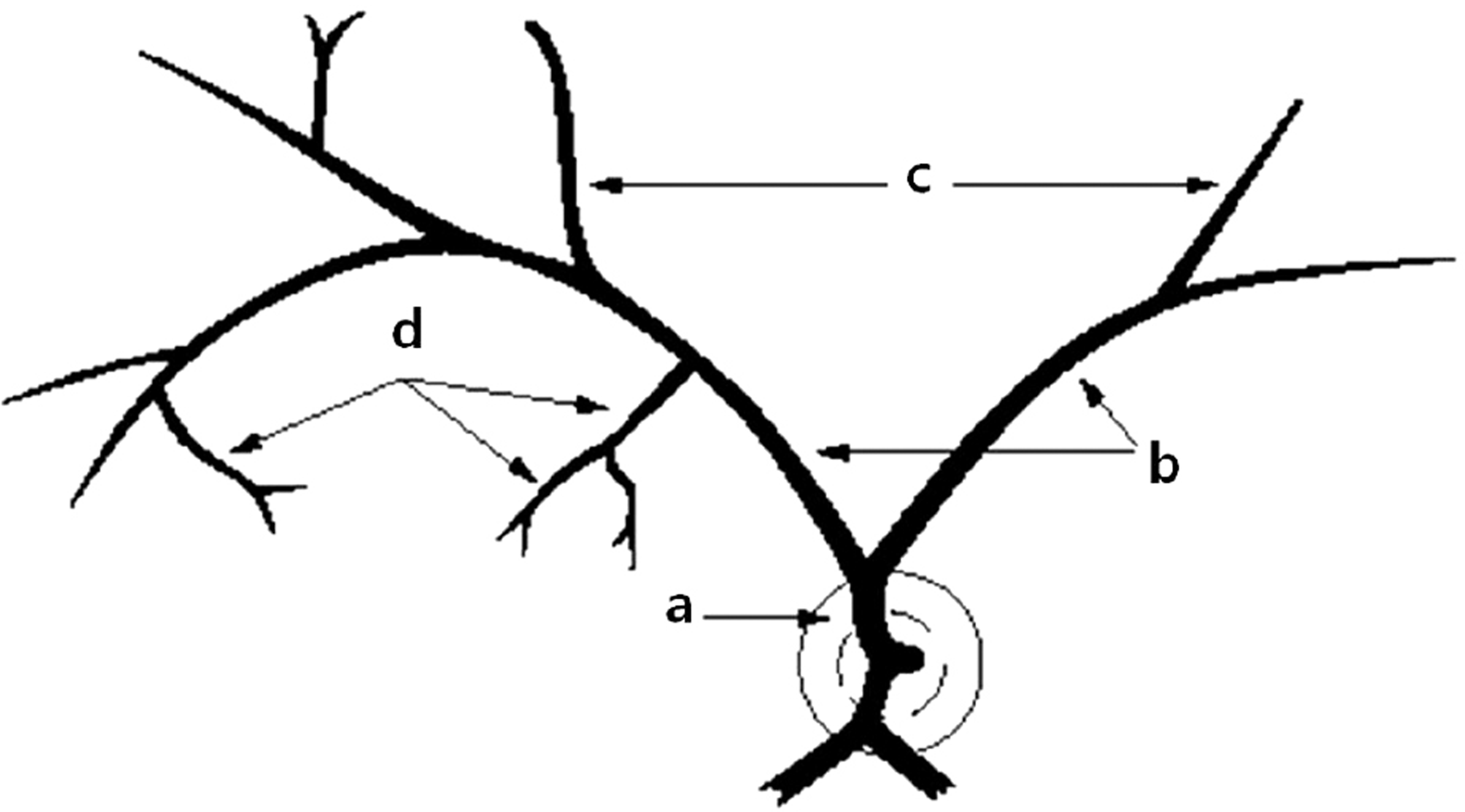
Figure 2.
Comparison of baseline visual acuity (A), final visual acuity (B), retinal ischemic area (C) and macular thickness (D) among the four groups which were divided by occlusion site in the branch retinal vein occlusion patients. DD = disc diameter; log MAR = logarithm of the minimal angle of resolution. * Statistical significance was found with ANOVA, Scheffe test (p < 0.05).
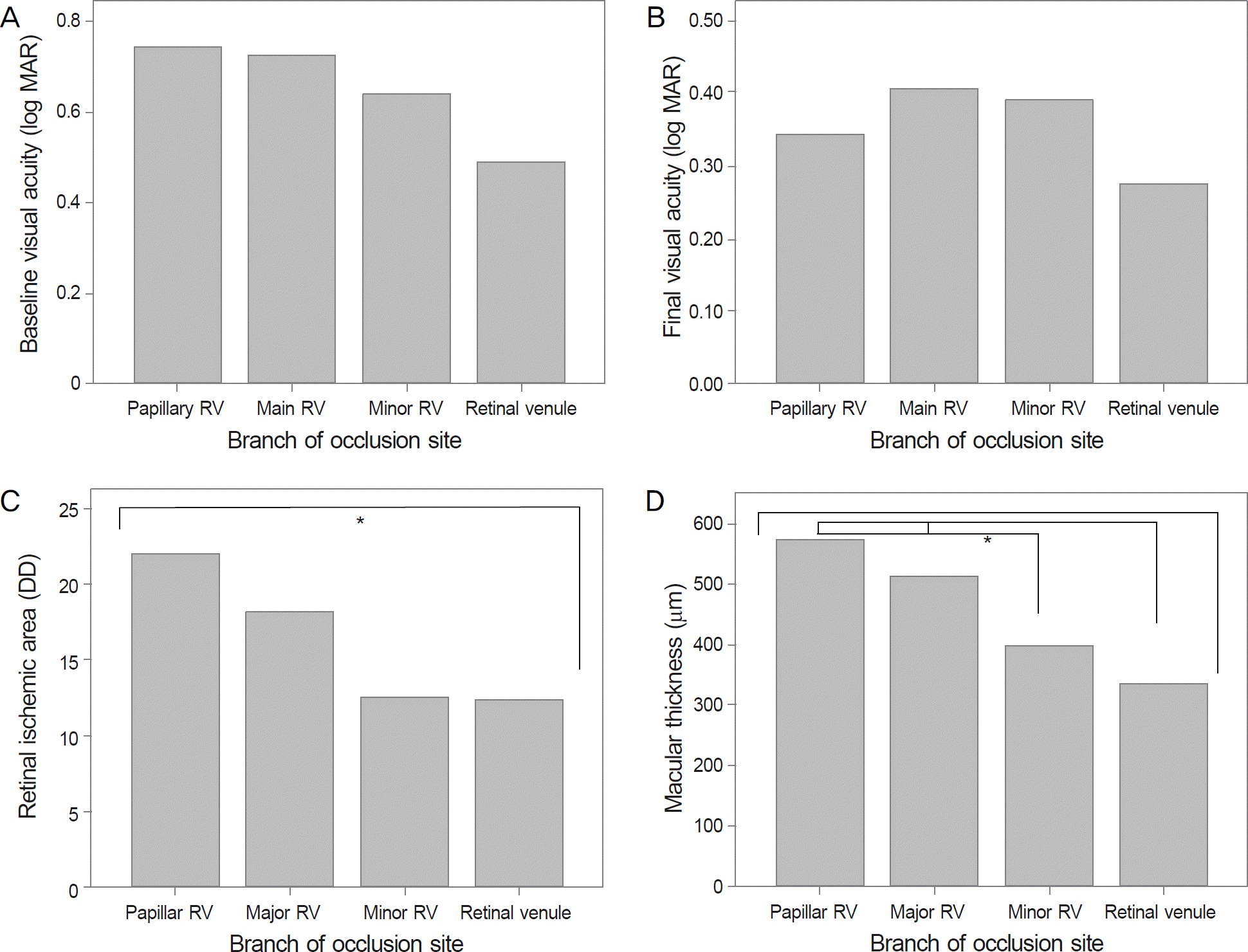
Table 1.
Characteristics of retinal vein occlusion patients
Table 2.
Characteristics of four groups which were divided by occlusion site in retinal vein occlusion subjects
| Characteristics | Papillary RV group (n = 15) | Main RV group (n = 34) | Minor RV group (n = 28) | Retinal venule group (n = 14) | p-value |
|---|---|---|---|---|---|
| Mean age (years) | 64.67 ± 6.80 | 59.42 ± 12.91 | 58.75 ± 11.40 | 60.07 ± 11.58 | 0.413* |
| Sex (M:F) | 4:11 | 14:20 | 11:17 | 3:11 | 0.497† |
| Laterality (R:L) | 7:8 | 18:16 | 12:16 | 6:8 | 0.857† |
| Location | 0.139† | ||||
| Superotemporal | 8 | 25 | 22 | 7 | |
| Inferotemporal | 7 | 9 | 6 | 7 | |
| Treatment | 0.424† | ||||
| IVB | 8 | 20 | 11 | 5 | |
| IVTA | 2 | 6 | 6 | 4 | |
| IVB + IVTA | 5 | 5 | 8 | 4 | |
| Baseline VA (log MAR) | 0.74 ± 0.40 | 0.72 ± 0.46 | 0.64 ± 0.53 | 0.49 ± 0.43 | 0.402* |
| Final VA (log MAR) | 0.34 ± 0.27 | 0.38 ± 0.65 | 0.39 ± 0.33 | 0.28 ± 0.21 | 0.625* |
| Mean IOP (mm Hg) | 14.20 ± 2.83 | 14.42 ± 3.49 | 14.54 ± 2.86 | 14.15 ± 2.61 | 0.977* |
| Macular thickness (μ m) | 575.33 ± 178.44 | 511.92 ± 218.02 | 397.21 ± 144.51 | 336.68 ± 120.55 | 0.001*,‡ |
Table 3.
Retinal ischemic area according to the occlusion site in branch retinal vein occlusion
| Papillary RV group (n = 15) | Main RV group (n = 34) | Minor RV group (n = 28) | Retinal venule group (n = 14) | p-value | |
|---|---|---|---|---|---|
| Retinal ischemic area (DD) | 22.00 ± 13.28 | 18.26 ± 10.12 | 12.52 ± 10.52 | 12.36 ± 11.92 | 0.025*,‡ |
| ≤5 DD | 1 | 3 | 8 | 6 | 0.002‡,‡ |
| 5-10 DD | 0 | 3 | 7 | 4 | |
| 10-20 DD | 9 | 19 | 9 | 0 | |
| 20 DD ≤ | 5 | 9 | 4 | 4 |
Table 4.
Perifoveal capillary ring state according to the occlusion site in branch retinal vein occlusion
| Papillary RV group (n = 15) | Main RV group (n = 34) | Minor RV group (n = 28) | Retinal venul grou (n = 14) | p p-value | |
|---|---|---|---|---|---|
| Intact | 10 | 14 | 17 | 8 | 0.286* |
| broken | 5 | 20 | 11 | 6 |
Table 5.
Macular ischemic area according to the occlusion site in branch retinal vein occlusion
| Papillary RV group (n = 15) | Main RV group (n = 34) | Minor RV group (n = 28) | Retinal venule grou (n = 14) | up p-value | |
|---|---|---|---|---|---|
| <90° | 1 | 3 | 7 | 1 | 0.080* |
| 90-180° | 7 | 6 | 8 | 7 | |
| 180-270° | 1 | 8 | 5 | 0 | |
| 270-360° | 6 | 17 | 8 | 6 |
 XML Download