Abstract
Purpose
To report a case of ischemic retinopathy due to suspicious gentamicin retinal toxicity after primary repair of a scleral laceration.
Case summary
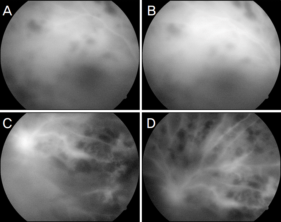
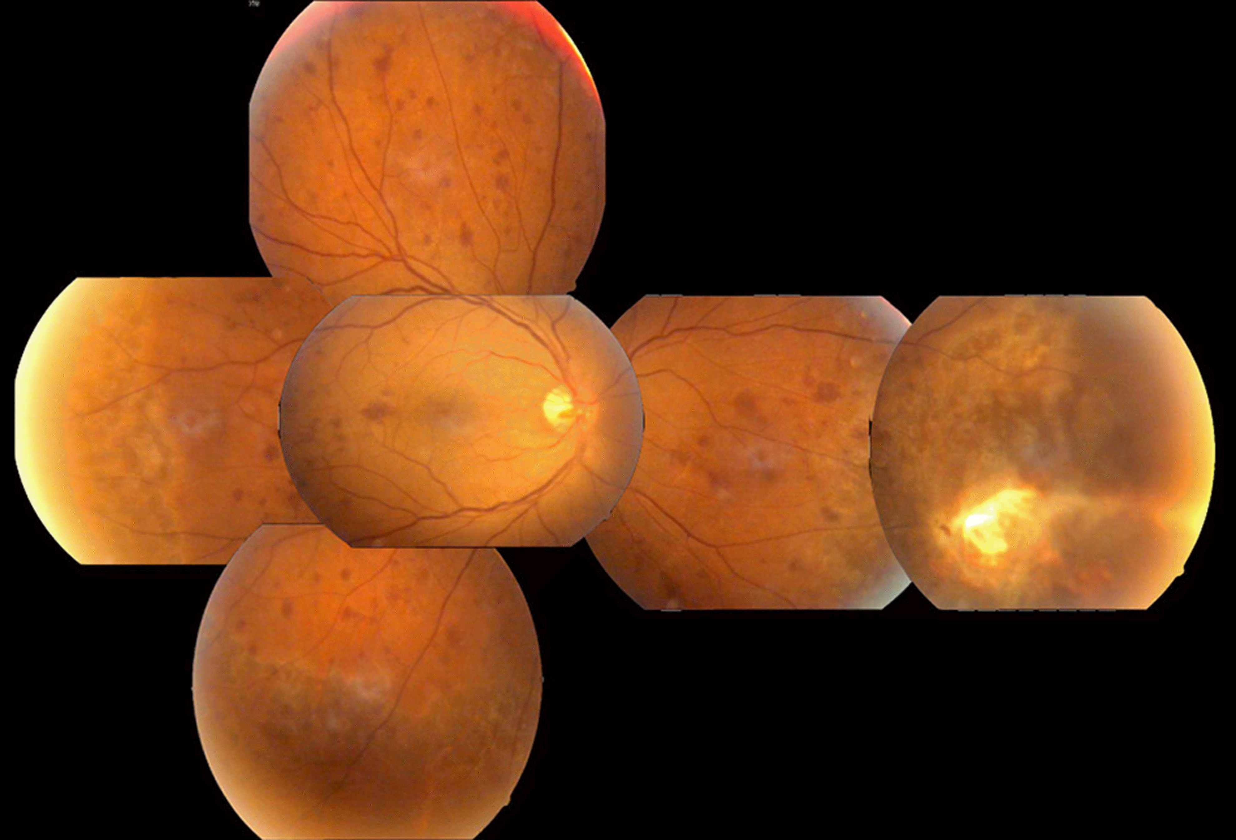
A 45-year-old man presented to our department with decreasing vision in his right eye after ocular trauma. Best corrected visual acuity (BCVA) was 0.02 in the right eye and slit lamp examination revealed scleral laceration. Both in-travenous and topical antibiotics (10% cefazolin and 2% gentamicin) were immediately administered. On intraoperative examination, a scleral laceration located 5 mm to 11 mm from nasal limbus, prolapsed vitreous body and partial division of medial rectus muscle were observed. After irrigation with gentamincin 0.2% around the wound, primary repair was performed. On postoperative day 3, fundus examination revealed a retinal break, barrier laser was performed. On post-operative day 4, diffuse retinal edema with intraretinal hemorrhage was observed as well as, superonasal ghost vessels. Subsequently, fluorescein angiography showed diffuse leakage of retinal vessels and a nonperfusion area at the periph-ery, especially on the nasal side. As vitreous opacity became worse, the patient underwent pars plana vitrectomy with endolaser. One month later, vitreous cavity was clearer and best visual acuity was 0.2.
Conclusions
Large doses of intraocular gentamicin ccan cause retinal toxicity. Increased gentamicin application through a scleral laceration may lead to toxic antibiotic levels. When a scleral laceration wound irrigation is performed, precautions are necessary to prevent retinal ischemia associated with gentamicin toxicity.
References
1. Balian JV. Accidental intraocular tobramycin injection: a case report. Ophthalmic Surg. 1983; 14:353–4.

2. Campochiaro PA, Conway BP. Aminoglycoside toxicity--a survey of retinal specialists. Implications for ocular use. Arch Ophthalmol. 1991; 109:946–50.

3. Campochiaro PA, LIM JI. Aminoglycoside toxicity in the treat-ment of endophthalmitis. The Aminoglycoside Toxicity Study Group. Arch Ophthalmol. 1994; 112:48–53.

4. D’Amico DJ, Caspers-Velu L, Libert J, et al. Comparative toxicity of intravitreal aminoglycoside antibiotics. Am J Ophthlmol. 1985; 100:264–75.
5. McDonald HR, Schatz H, Allen AW, et al. Retinal toxicity secon-dary to intraocular gentamicin. Ophthalmlogy. 1986; 93:871–7.
6. Chung DJ, Choi SH, Kim SD. Two cases of ischemic retinopathy due to intravitreal gentamicin toxicity after vitrectomy. J Korean Ophthalmol. 1993; 34:1183–7.
7. Loewenstein A, Zemel E, Vered Y, et al. Retinal toxicity of genta-micin after subconjunctival injection performed adjacent to thin-ned sclera. Ophthalmology. 2001; 108:759–64.

8. Vakulenko SB, Mobashery S. Versatility of aminoglycosides and prospects for their future. Clin Micobiol Rev. 2003; 16:430–50.

9. Conway BP, Campochiaro PA. Macular infarction after endoph-thalmitis treated with vitrectomy and intravitreal gentamicin. Arch Ophthalmol. 1986; 104:367–71.

10. Snider JD 3rd, Cohen HB, Chenoweth RG. Acute ischemic retin-opathy secondary to intraocular injection of gentamicin. Ryan SJ, Dawson AK, Little HL, editors. Retinal Disease. Orlando, FL: Grune and Stratton;1985. p. chap. 39.
11. D’Amico DJ, Libert J, Kenyon KR, et al. Retinal toxicity of intra-vitreal gentamicin. An electron microscopic study. Invest Ophthalmol Vis Sci. 1984; 25:564–72.
12. Peyman GA, Paque JT, Meisels HI, Bennett TO. Postoperative en-dophthalmitis: a comparison of methods for treatment and prophy-laxis with gentamicin. Ophthalmic Surg. 1975; 6:45–55.
Figure 1.
(A) A photograph of the right eye before primary repair shows a full-thickness scleral laceration, partial division of medial rectus muscle and prolapsed vitreous body. (B) Irrigation with 10 ml of gentamicin 0.2% was performed around sclera laceration.

Figure 2.
Fundus photograph taken 3 days later primay repair of sclera laceration showing retinal edema, intraretinal hemor-rhage, and ghost vessels.


 PDF
PDF ePub
ePub Citation
 Print
Print


 XML Download