

 PDF
PDF ePub
ePub Citation
 Print
Print


Abstract
Case summary
A 69-year-old man visited the hospital with visual disturbance in both eyes. His mother had been diag-nosed with rubella during pregnancy, exhibiting typical fever and rashes. His visual acuity and hearing ability had been poor since birth. Corrected visual acuity was 0.3 in the right eye and 0.4 in the left eye. Slit lamp examination revealed bi-lateral zonular cataracts. On pure tone audiometry test, pure tone hearing threshold was 73 dB in the right ear and 72 dB in the left ear, corresponding to severe hearing loss in both ears. Echocardiogram showed a 1.5 cm-sized ostium se-cundum atrial septal defect, causing atrial fibrillation. Laboratory workup revealed an extremely high level of IgG antibody (titer = 1:301), and negative IgM antibody. A rubella IgG avidity test was 95.5%, suggesting remote rubella infection. Chromosomal analysis from peripheral blood did not show any abnormalities. The patient was diagnosed with congenital rubella syndrome with bilateral zonular cataracts. Two months after cataract surgery on both eyes, visual acuity steadily improved to 1.0 in both eyes.
Conclusions
Congenital rubella syndrome is comprised of physical abnormalities such as sensorineural hearing loss, eye defects including congenital cataract, and cardiovascular defects due to gestational rubella infection. The possibility of congenital rubella syndrome should be considered even in old age, and a systemic multi-organ approach is necessary for therapeutic planning.
References
1. Duszak RS. Congenital rubella syndrome-major review. Optometry. 2009; 80:36–43.

2. Krause AC. Congenital cataracts following rubella in pregnancy. Ann Surg. 1945; 122:1049–55.
3. Webster WS. Teratogen update: congenital rubella. Teratology. 1998; 58:13–23.
4. Shin YJ, Choi BY, Park HB, et al. Cost-benefit analysis on rubella vaccination policy. Korean J Prev Med. 1994; 27:337–65.
5. Choe YJ, Hu JK, Song KM, et al. Evaluation of an expanded case definition for vaccine-modified measles in a school outbreak in South Korea in 2010. Jpn J Infect Dis. 2012; 65:371–5.
6. Kim SS, Han HW, Go U, Chung HW. Sero-epidemiology of mea-sles and mumps in Korea: impact of the catch-up campaign on measles immunity. Vaccine. 2004; 23:290–7.
7. Lee KY, Sul JH, Lee YN, et al. A measurement of rubella anti-bodies among Korean children by enzyme-linked immunosorbent assay. Yonsei Med J. 1981; 22:41–8.
8. Chi JG, Suh EH, Lee K. Congenital rubella syndrome with necrotiz-ing panencephalitis (an autopsy case). Seoul J Med. 1987; 28:275–9.
9. O'Neill JF. The ocular manifestations of congenital infection: a study of the early effect and long-term outcome of maternally transmitted rubella and toxoplasmosis. Trans Am Ophthalmol Soc. 1998; 96:813–79.
10. Hanna C. Embryopathic time-table of cataract formation in con-genital rubella. J Pediatr Ophthalmol. 1976; 13:266–73.
11. Khokhar S, Agarwal T, Kumar G, et al. Lenticular abnormalities in children. J Pediatr Ophthalmol Strabismus. 2012; 49:32–7.
12. Gerber SL, Helveston EM. Subretinal neovascularization in a 10-year-old child. J Pediatr Ophthalmol Strabismus. 1992; 29:250–1.
13. Arnold JJ, McIntosh ED, Martin FJ, Menser MA. A fifty-year fol-low-up of ocular defects in congenital rubella: late ocular manifestations. Aust N Z J Ophthalmol. 1994; 22:1–6.
14. Givens KT, Lee DA, Jones T, Ilstrup DM. Congenital rubella syn-drome: ophthalmic manifestations and associated systemic disorders. Br J Ophthalmol. 1993; 77:358–63.
15. Kobayashi S, Hamatsu Y, Kimura R, et al. A case of bilateral con-genital cataract left untreated for 60 years. J Eye. 2005; 22:81–5.
16. Liu Y, Guo J, Zhao RF, Wang L. Unilateral pulmonary artery steno-sis and late-onset cataract in an adult: a case of suspected con-genital rubella syndrome. Chin Med J. 2012; 125:549–51.
Figure 1.
Bilateral zonular cataract of the patient. Haze in lamellar zone between the cortex and nucleus was clearly demarcated un-der slit view illumination. Multiple speckle opacities in the nucleus, and a clear zone in the out cortex are also characteristic features of cataract in congenital rubella syndrome.

Figure 2.
Pure tone audiometry of the patient. Hearing threshold was 73 dB in the right ear, and was 72 dB in the left ear. Air con-duction was poorer than bone conduction by more than 10 dB, and the bond conduction threshold was lesser than 25 dB in both ears. Mixed form of conduction and sensorineural hearing loss was diagnosed, and the degree of hearing loss was categorized as severe grade in both ears.

Figure 3.
Two dimensional echocardiogram of the patient. There was a 1.5 cm sized ostium secundum atrial septal defect, causing left to right cardiac shunt, pulmonary hypertension, and atrial fibrillation of the patient.
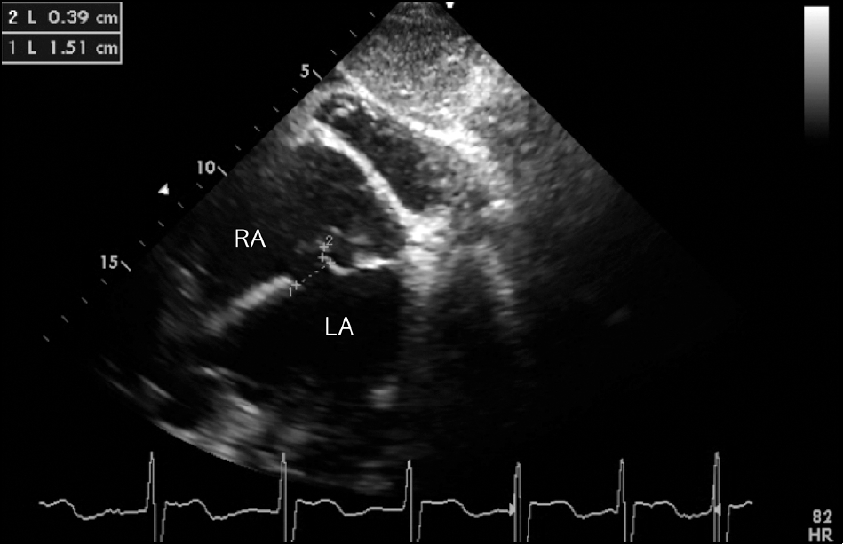
Figure 4.
Postoperative red-free fundus photos of the patient. Superotemporal retinal nerve fiber layer thinning was pointed with ar-rows in the right eye.
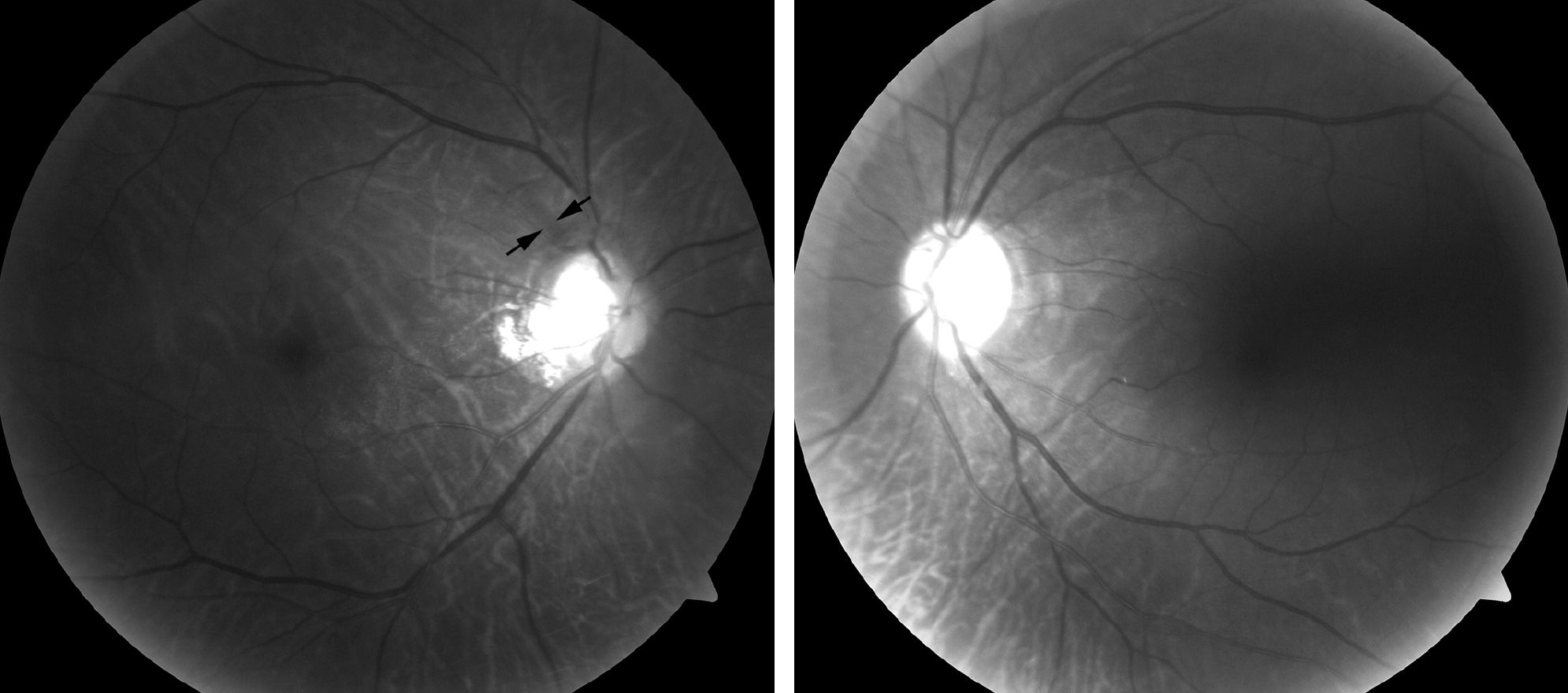
Figure 5.
Retinal nerve fiber layer (RNFL) analysis of the patient by spectral domain opitcal coherence to-mography (OCT) (Carl Zeiss Meditec, Germany). Superotemporal RNFL thinning was observed in the right eye. Slight inferonasal RNFL thinning was also noted in RNFL clock hour analysis, but it was not evi-dent in RNFL deviation map.

 XML Download