Abstract
Case summary
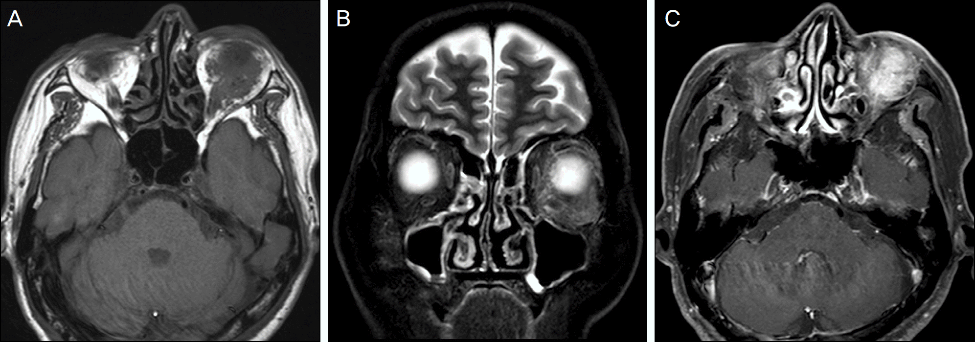
A 61-year-old male visited our clinic with diplopia, which had developed approximately 5 months earlier. Magnetic resonance imaging of the orbit showed an ill-defined well-enhanced mass in the left inferior orbit. Incisional biop-sy of the orbital mass was performed. Histopathological examination revealed diffuse deposits of an amorphous, eosino-philic substance in the extracellular matrix and vessels with lymphocytes infiltration. Lymphocytes were positive for the im-munohistochemical stain against the CD20 and κ-light chain antigens. The amorphous material stained positive for κ-light chain antigen, and Congo red staining showed birefringence.
References
1. Spencer WH. Ophthalmic Pathology, 3rd ed. Philadelphia:: WB Saunders;1985. p. 2672–98.
2. Yoon JS, Ma KT, Kim SJ, et al. Prognosis for patients in a Korean population with ocular adnexal lymphoproliferative lesions. Ophthal Plast Reconstr Surg. 2007; 23:94–9.

3. Meunier J, Lumbroso-Le Rouic L, Vincent-Salomon A, et al. Ophthalmologic and intraocular non-Hodgkin's lymphoma: a large single centre study of initial characteristics, natural history, and prognostic factors. Hematol Oncol. 2004; 22:143–58.

4. Knowles DM 2nd, Jakobiec FA, Rosen M, Howard G. Amyloidosis of the orbit and adnexae. Surv Ophthalmol. 1975; 19:367–84.
5. Picken MM. New insights into systemic amyloidosis: the im-portance of diagnosis of specific type. Curr Opin Nephrol Hypertens. 2007; 16:196–203.

6. Leibovitch I, Selva D, Goldberg RA, et al. Periocular and orbital amyloidosis: clinical characteristics, management, and outcome. Ophthalmology. 2006; 113:1657–64.
7. Murdoch IE, Sullivan TJ, Moseley I, et al. Primary localised amy-loidosis of the orbit. Br J Ophthalmol. 1996; 80:1083–6.

8. Goshe JM, Schoenfield L, Emch T, Singh AD. Myeloma-asso-ciated orbital amyloidosis. Orbit. 2010; 29:274–7.

9. Dacic S, Colby TV, Yousem SA. Nodular amyloidoma and primary pulmonary lymphoma with amyloid production: a differential di-agnostic problem. Mod Pathol. 2000; 13:934–40.

10. Caulet S, Robert I, Bardaxoglou E, et al. Malignant lymphoma of mucosa associated lymphoid tissue: a new etiology of amyloidosis. Pathol Res Pract. 1995; 191:1203–7.

11. Goteri G, Ranaldi R, Pileri SA, Bearzi I. Localized amyloidosis and gastrointestinal lymphoma: a rare association. Histopathology. 1998; 32:348–55.

12. Wieker K, Röcken C, Koenigsmann M, et al. Pulmonary low-grade MALT-lymphoma associated with localized pulmonary amyloidosis. A case report. Amyloid. 2002; 9:190–3.
Figure 2.
(A) T1-weighted precontrast axial image demonstrating an ill-defined mass in the left orbit isointense to extraocular muscle. (B) T2-weighted precontrast coronal image showing a mass with subtle high signal intensity in the left inferior orbit. (C) T1-weighted postcontrast image demonstrates a well-enhancing soft tissue mass.

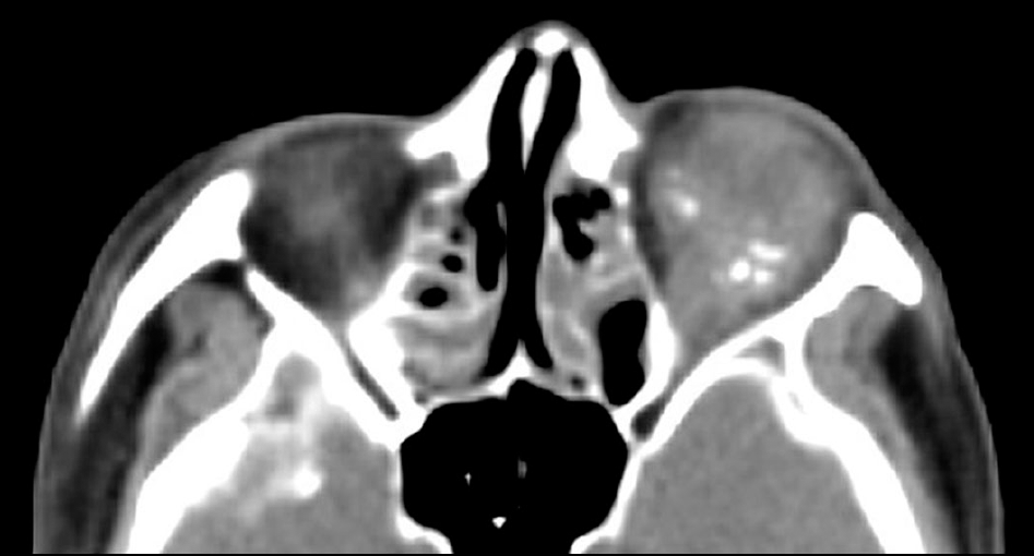
Figure 3.
Axial nonenhanced CT scan reveals left inferior or-bital soft tissue mass with calcifications.

Figure 4.
(A) Histopathologic examination reveals diffuse deposits of an amorphous, eosinophilic substance in the extracellular ma-trix and vessels with lymphocytes infiltration (hematoxylin and eosin, ×200). (B) High-power magnification shows that the cells are predominantly small lymphocytes (hematoxylin and eosin, ×400). (C) Lymphocytes stain positive for the immunohistochemical stain against the CD20 antigen (×400). (D) Lymphocytes and an amorphous material stain positive for the immunohistochemical stain against κ-light chain antigen (×100). (E) The amorphous material stains positive with Congo red and F, demonstrates ap-ple-green birefringence under the polarized light (Congo red, ×100).


 PDF
PDF ePub
ePub Citation
 Print
Print


 XML Download