Abstract
Purpose
We investigated the success rate of surgery and binocular function after surgery in intermittent exotropia with good preoperative binocular function.
Methods
Thirty-eight intermittent exotropia patients who had good stereopsis of 40 seconds according to the Titmus test, showed fusion by Worth-4-dot test preoperatively, and had at least 1 year of postoperative follow-up were included in the present study. The age at operation, angle of exodeviation, visual acuity, stereopsis with Titmus test and fusional status with Worth-4-dot test after surgery were analyzed. A surgical success was defined as postoperative angle of deviation less than 10 prism diopter (PD).
Results
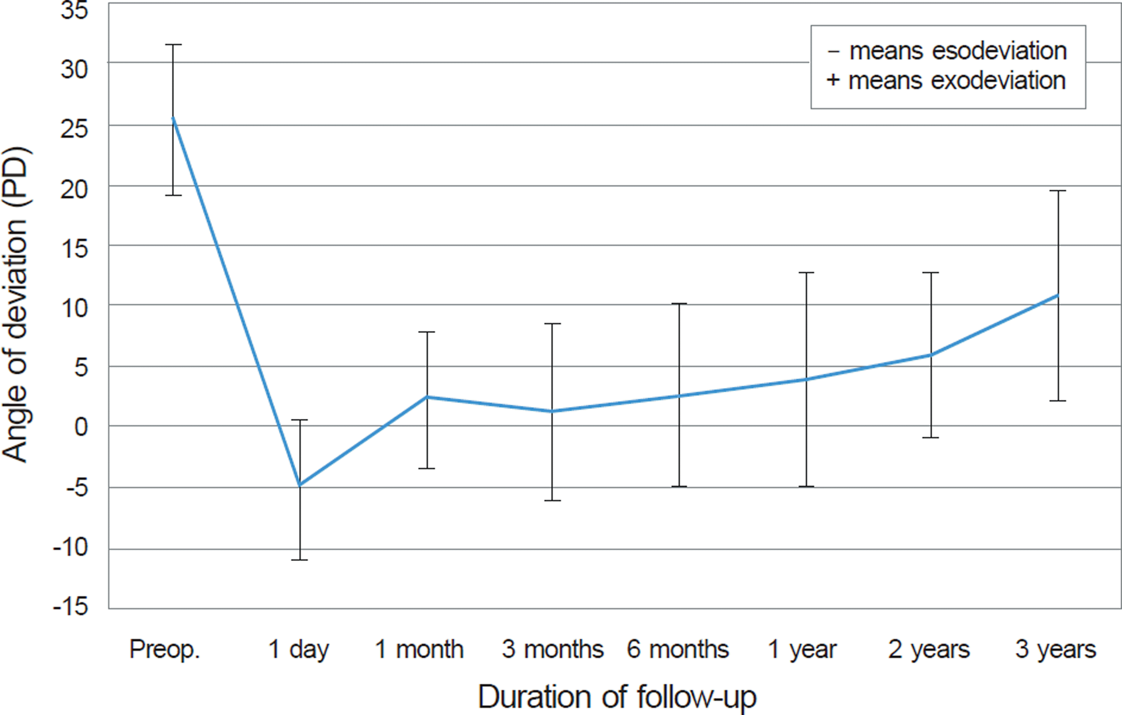
The patient mean age at the time of the operation was 7.9 years. The mean preoperative angle of exodeviation was 25.5 PD at far distance and 27.5 PD at near distance. The mean follow-up time was 22.9 months. The success rate of surgery was 81.6% at 6 months, 68.4% at 1 year and 60.5% on the last visit. Seventeen patients (44.7%) had stereopsis of 40 seconds and showed fusion at far and near distance after surgery. The stereopsis was worse than 100 seconds in 2 patients (5.3%), and fusion was maintained at only near distance in 15 patients (39.5%). In 7 patients (18.4%), the stereo-psis decreased to 200 seconds or worse, or there was no fusion after surgery.
Conclusions
The recurrence of exodeviation was a major cause of the surgical failure in the intermittent exotropia with good preoperative binocular function. Moreover, binocular function may decrease postoperatively in intermittent exotropia with good preoperative binocular function, so careful follow-up may be required to maintain this function.
Go to : 
References
1. Jenkins R. Demographics: geographic variations in the prevalence and management of exotropia. Am Orthopt J. 1992; 42:82–7.

2. Figueira EC, Hing S. Intermittent exotropia: comparison of treatments. Clin Experiment Ophthalmol. 2006; 34:245–51.

3. von Noorden GK. Binocular vision and ocular motility: Theory and management of strabismus, 5th ed. St. Louis:: CV Mosby;1996. p. 341–59.
4. Kushner BJ, Morton GV. Postoperative binocularity in adults with longstanding strabismus. Ophthalmology. 1992; 99:316–9.

5. Heo NH, Paik HJ. The relationship between binocular function and the surgical outcome of intermittent exotropia. J Korean Ophthalmol Soc. 2001; 42:1588–93.
6. Wright KW, Ryan SJ. Color atlas of ophthalmic surgery: stra-bismus. Philadelphia:: Lippincott;1991. p. 241–3.
7. Burian HM. Exodeviation: their classification, diagnosis and treatment. Am J Ophthalmol. 1966; 62:1161–6.
8. Burian HM, Franceschetti AT. Evaluation of diagnostic methods for the classification of exodeviations. Am J Ophthalmol. 1971; 1:34–41.

9. Burian HM, Spivey BE. The surgical management of exodeviations. Am J Ophthalmol. 1965; 59:603–20.
10. Shin YJ, Chang BL. The clinical outcome of the consecutive eso-tropia after surgical correction. J Korean Ophthalmol Soc. 2003; 44:2085–90.
11. Edelman PM, Brown MH, Murphree AL, Wright KW. Consecutive esodeviation··· then what? Am Orthopt J. 1988; 38:111–6.
12. Campos EC, von Noorden GK. Binocular vision and ocular mo-tility; Theory and management of strabismus, 6th ed. St. Louis: Mosby;2002. p. chap 13–17.
13. Suh WJ, Lee UK, Kim MM. Change of postoperative distance ster-eoacuity in intermittent exotropic patients. J Korean Ophthalmol Soc. 2000; 41:758–63.
14. Lee SY, Kim SJ, Ahn JH, et al. The effects of surgery on binocular function in intermittent exotropia. J Korean Ophthalmol Soc. 1999; 40:3180–6.
15. Rutstein RP, Daum KM. Anomalies of binocular vision: Diagnosis and management. 1st ed. St. Louis:: Mosby;1997. p. 111–46.
16. Lee SY. Comparison of distance and near stereoacuity in normal and intermittent exotropic children. J Korean Ophthalmol Soc. 2001; 42:624–9.
17. Beneish R, Flanders M. The role of stereopsis and early post-operative alignment in long-term surgical results of intermittent exotropia. Can J Ophthalmol. 1994; 29:119–24.
18. Gill MK, Drummond GT. Indications and outcomes of strabismus repair in visually mature patients. Can J Ophthalmol. 1997; 32:436–40.
19. Rosenbaum AL, Santiago AP. Clinical strabismus management: Principles and surgical techniques, 1st ed. Philadelphia:: Saunders;1999. p. 156–68.
21. Yildirim C, Mutlu FM, Chen Y, Altinsoy HI. Assessment of central and peripheral fusion and near and distance stereoacuity in inter-mittent exotropic patients before and after strabismus surgery. Am J Ophthalmol. 1999; 128:222–30.

22. Pratt-Johnson JA, Barlow JM, Tillson G. Early surgery for inter-mittent exotropia. Am J Ophthalmol. 1977; 84:689–94.
Go to : 
 | Figure 2.Change of the angle of deviation during the follow-up after surgery for intermittent exotropia. |
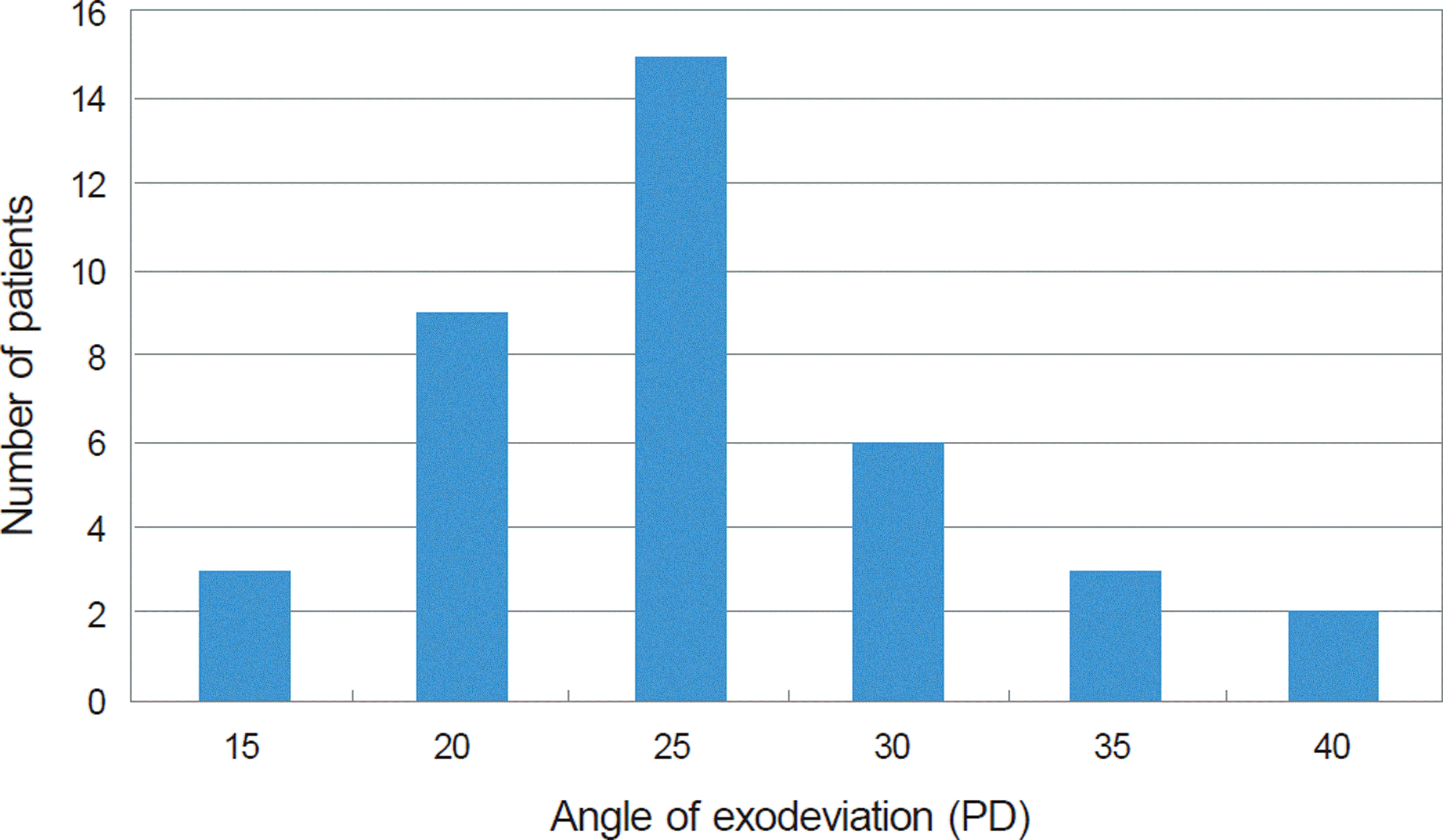
 | Figure 3.Postoperative streopsis (A) and fusion (B). * Fusion at far and near; † Fusion at near; ‡ No fusion. |
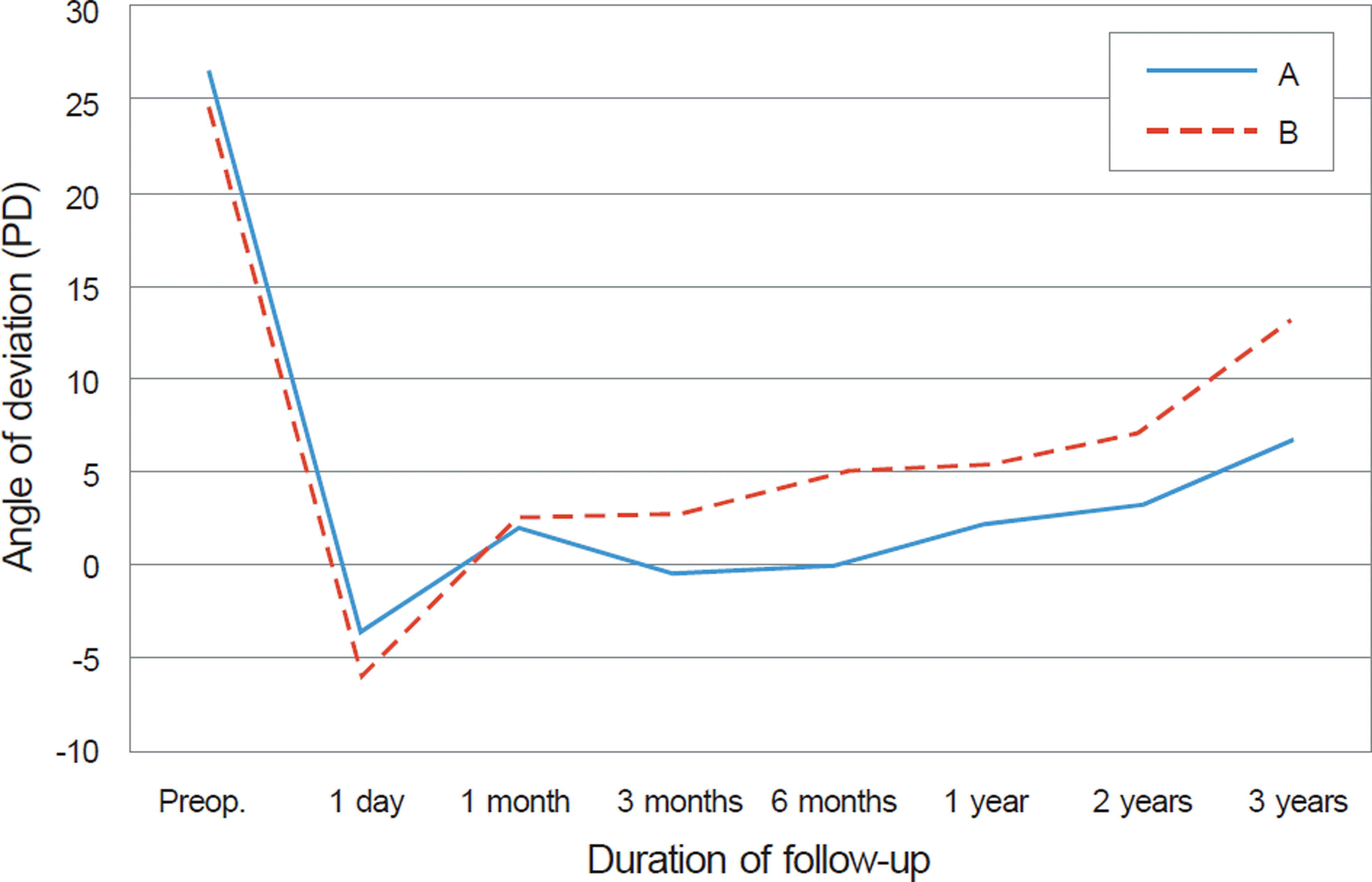
 | Figure 4.Change of the postoperative angle of deviation be-tween maintained binocular vision group and reduced binoc-ular vision group. (A) Patients who maintained preoperative good binocular function after surgery (n = 17). (B) patients who decreased preoperative binocular function after surgery (n = 21). |
Table 1.
The characteristics of patients with intermittent exo-tropia
Table 2.
Comparison of preoperative and postoperative data between maintained binocular vision group and reduced binocular vi-sion group
| Maintained binocular vision group (n = 17) | Reduced binocular vision group (n = 21) | p-value | |
|---|---|---|---|
| Sex (M:F) | 6 : 11 | 9 : 12 | 0.74* |
| Age at surgery (years, range) | 8.4 ± 2.2 (5 to 13) | 7.4 ±1.7 (5 to 9) | 0.13† |
| Follow-up (months, range) | 19.1 ± 9.5 (12 to 36) | 26.0 ± 12.5 (12 to 48) | 0.07† |
| Preoperative angle of exodeviation (PD, range) | |||
| Far | 26.5 ± 6.8 (15 to 40) | 24.6 ± 5.7 (15 to 40) | 0.37† |
| Near | 28.5 ± 6.3 (20 to 40) | 26.7 ± 5.5 (15 to 43) | 0.34† |
| Postoperative angle of deviation at last visit (PD, range) | |||
| Far | 3.1 ± 11.1 (-30 to 20) | 8.9 ± 10.1 (-10 to 25) | 0.10† |
| Near | 3.5 ± 11.1 (-30 to 20) | 9.9 ± 11.1 (-10 to 30) | 0.09† |
| Postoperative patching therapy (No.) | 8 | 9 | 0.51* |
| Duration of postoperative patching therapy (weeks, range) | 1.8 ± 1.8 (1 to 6) | 1.8 ± 0.9 (1 to 4) | 0.31‡ |
| Result at last visit (No.) | |||
| Surgical success | 12 | 11 | |
| Overcorrection | 1 | 0 | 0.20* |
| Recurrence | 4 | 10 |


 PDF
PDF ePub
ePub Citation
 Print
Print


 XML Download