Abstract
Purpose
To evaluate the benefits of one day, one eye ICL implantation which allows the ICL size of the later-operated eye to be adjusted after evaluating the postoperative vault of the first-operated eye in order to reduce the postoperative ICL size exchange rates of both eyes.
Methods
A total of 426 eyes of 213 patients who received one day, one eye bilateral ICL implantation were included in the present study. The cases where a different ICL size was implanted in the later-operated eye because of high or low post-operative vault of the first-operated eye were analyzed as well as the ICL exchange rates.
Results
Among 213 patients, same size ICLs were implanted in both eyes in 188 patients (88%) as planned. However, a different ICL size was implanted in the later-operated eye in 25 patients (12%). Eight eyes of 8 patients out of 25 patients needed their ICL size exchanged during the follow-up period and all 8 eyes were first-operated eyes. This occurred in 8 pa-tients (3.8%) out of 213 patients.
Go to : 
References
1. Han SY, Lee KH. Long term effect of ICL implantation to treat high myopia. J Korean Ophthalmol Soc. 2007; 48:465–72.
2. Han SY, Moon SJ, Kim HS, et al. Intraindividual comparison of ICL and toric ICL implantation in the correction of high myopia with astigmatism. J Korean Ophthalmol Soc. 2010; 51:802–8.

3. Du GP, Huang YF, Wang LQ, et al. [Outcome after treatment of myopia with implantable collamer lens]. Zhonghua Yan Ke Za Zhi. 2011; 47:146–50.
4. Pitault G, Leboeuf C, Leroux Les Jardins S, et al. [Ultrasound bio-microscopy of posterior chamber phakic intraocular lenses: a com-parative study between ICL and PRL models]. J Fr Ophtalmol. 2005; 28:914–23.
5. Alfonso JF, Lisa C, Abdelhamid A, et al. Posterior chamber phakic intraocular lenses after penetrating keratoplasty. J Cataract Refract Surg. 2009; 35:1166–73.

6. Kamiya K, Shimizu K, Igarashi A, et al. Clinical evaluation of opti-cal quality and intraocular scattering after posterior chamber phak-ic intraocular lens implantation. Invest Ophthalmol Vis Sci. 2012; 53:3161–6.

7. Kamiya K, Igarashi A, Shimizu K, et al. Visual performance after posterior chamber phakic intraocular lens implantation and wave-front-guided laser in situ keratomileusis for low to moderate myopia. Am J Ophthalmol. 2012; 153:1178–86.e1.

8. Hasegawa A, Kojima T, Isogai N, et al. Astigmatism correction: Laser in situ keratomileusis versus posterior chamber collagen co-polymer toric phakic intraocular lens implantation. J Cataract Refract Surg. 2012; 38:574–81.

9. Portaliou DM, Kymionis GD, Panagopoulou SI, et al. Long-term results of phakic refractive lens implantation in eyes with high myopia. J Refract Surg. 2011; 27:787–91.

10. Alfonso JF, Baamonde B, Fernández-Vega L, et al. Posterior cham-ber collagen copolymer phakic intraocular lenses to correct my-opia: five-year follow-up. J Cataract Refract Surg. 2011; 37:873–80.

11. Chung YW, Byun YS, Chung SK. Long-term changes in tilt, de-centration and anterior chamber depth after implantable collamer lens imsertion. J Korean Ophthalmol Soc. 2011; 52:157–62.
12. Yoon JM, Moon SJ, Lee KH. Clinical outcomes of toric implant-able collamer lens implantation. J Korean Ophthalmol Soc. 2009; 50:839–51.

13. Fernandes P, González-Méijome JM, Madrid-Costa D, et al. Implantable collamer posterior chamber intraocular lenses: a re-view of potential complications. J Refract Surg. 2011; 27:765–76.

14. Gonvers M, Othenin-Girard P, Bornet C, Sickenberg M. Implantable contact lens for moderate to high myopia: short-term follow-up of 2 models. J Cataract Refract Surg. 2001; 27:380–8.
15. Sanders DR, Vukich JA. ICL in Treatment of Myopia (ITM) Study Group. Incidence of lens opacities and clinically significant cata-racts with the implantable contact lens: comparison of two lens designs. J Refract Surg. 2002; 18:673–82.
16. Kojima T, Maeda M, Yoshida Y, et al. Posterior chamber phakic implantable collamer lens: changes in vault during 1 year. J Refract Surg. 2010; 26:327–32.

17. Alfonso JF, Lisa C, Abdelhamid A, et al. Three-year follow-up of subjective vault following myopic implantable collamer lens implantation. Graefes Arch Clin Exp Ophthalmol. 2010; 248:1827–35.

18. Du GP, Huang YF, Wang LQ, et al. Changes in objective vault and effect on vision outcomes after implantable Collamer lens im-plantation: 1-year follow-up. Eur J Ophthalmol. 2012; 22:153–60.

19. Lege BA, Haigis W, Neuhann TF, Bauer MH. Age-related behavior of posterior chamber lenses in myopic phakic eyes during accom-modation measured by anterior segment partial coherence inter- ferometry. J Cataract Refract Surg. 2006; 32:999–1006.
20. Choi KH, Chung SE, Chung TY, Chung ES. Ultrasound biomicro-scopy for determining visian implantable contact lens length in phakic IOL implantation. J Refract Surg. 2007; 23:362–7.

21. Lee DH, Choi SH, Chung ES, Chung TY. Correlation between pre-operative biometry and posterior chamber phakic visian implant-able collamer lens vaulting. Ophthalmology. 2012; 119:272–7.

22. Seo JH, Kim MK, Wee WR, Lee JH. Effects of white-to-white di-ameter and anterior chamber depth on implantable collamer lens vault and visual outcome. J Refract Surg. 2009; 25:730–8.

23. Bechmann M, Ullrich S, Thiel MJ, et al. Imaging of posterior chamber phakic intraocular lens by optical coherence tomography. J Cataract Refract Surg. 2002; 28:360–3.

24. Alfonso JF, Lisa C, Palacios A, et al. Objective vs subjective vault measurement after myopic implantable collamer lens implantation. Am J Ophthalmol. 2009; 147:978–83.e1.

25. Kojima T, Yokoyama S, Ito M, et al. Optimization of an implant-able collamer lens sizing method using high-frequency ultrasound biomicroscopy. Am J Ophthalmol. 2012; 153:632–7.

26. Yokoyama S, Kojima T, Horai R, et al. Repeatability of the ciliary sulcus-to-sulcus diameter measurement using wide-scanning-field ultrasound biomicroscopy. J Cataract Refract Surg. 2011; 37:1251–6.

Go to : 
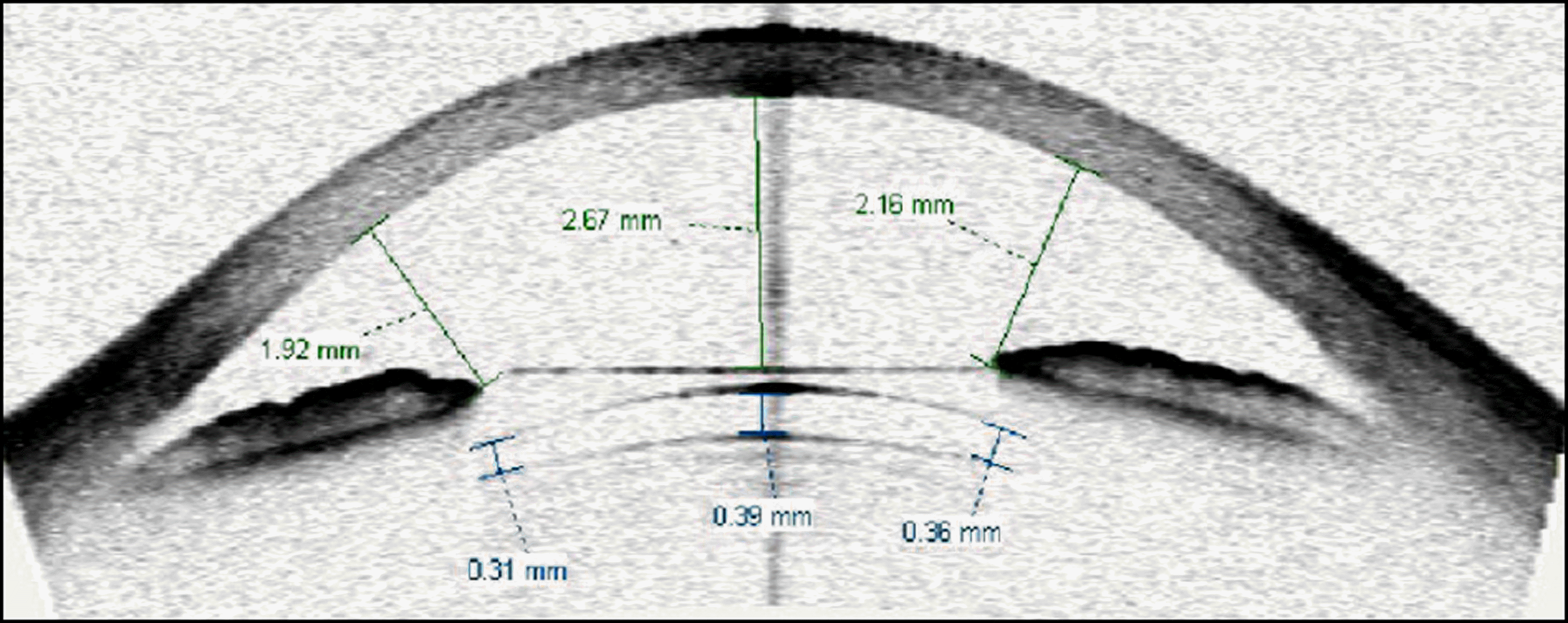
 | Figure 1.Visante OCT image of patient who received ICL implantation. The postoperative vault of this patient is 0.39 mm. |
Table 1.
Basic characteristics of 426 eyes of 213 patients wh received bilateral ICL implantation
Table 2.
The change of ICL sizes from first-operated eye to later-operated eye in 25 patients in which different size ICLs were implanted
| ICL size (mm) (first-operated eye ? later-operated eye) | Number of patients |
|---|---|
| 115 ? 120 | 7 |
| 120 ? 125 | 4 |
| 125 ? 130 | 0 |
| 130 ? 125 | 0 |
| 125 ? 120 | 8 |
| 120 ? 115 | 6 |
| Total | 25 |
Table 3.
The postoperative results of two groups which are first-operated eyes and later-operated eyes in 213 patients (426 eyes)
| First-opearted eyes | Later-operated eyes | p-value | |
|---|---|---|---|
| Implanted ICL size (mm) | 11.9 ± 0.33 | 11.9 ± 0.32 | 0.83* |
| Postoperative vault (mm) | 0.47 ± 0.24 | 0.46 ± 0.21 | 0.65* |
| Low vault (eyes) | 35 (16%) | 36 (17%) | 1.00† |
| Ideal vault (eyes) | 159 (75%) | 158 (74%) | 1.00† |
| High vault (eyes) | 19 (9%) | 19 (9%) | 1.00† |
| ICL size exchanged (eyes) | 10 (4.7%) | 4 (1.9%) | 0.17† |
Table 4.
The analysis of ICL size in 14 eyes in which ICL size exchange was done
Table 5.
The ICL exchange rate according to the size of implanted ICL


 PDF
PDF ePub
ePub Citation
 Print
Print


 XML Download