

 PDF
PDF ePub
ePub Citation
 Print
Print


Abstract
Purpose
To investigate the possibility of blepharoptosis as a complication after panretinal photocoagulation using oph-thalmoscopic contact lens.
Methods
We prospectively evaluated patients who were diagnosed with diabetic retinopathy and scheduled to be treated with panretinal photocoagulation. Margin reflex distance 1 (MRD1), levator function, palpebral fissure height and width, and tarsal plate height were measured at the day of photocoagulation and 3 months after treatment.
Results
MRD1 was decreased in 8 eyes (25.8%), levator function was decreased in 5 eyes (16.1%), and palpebral fissure height was decreased in 6 eyes (19.4%). The decrement of MRD1 and palpebral fissure height after photocoagulation were significant ( p = 0.008, p = 0.031, respectively). There was a significant negative correlation between MRD1 decre-ment and tarsal plate height ( p = 0.045).
Go to : 
References
1. The Diabetic Retinopathy Study Research Group. Photocoagulation treatment of proliferative diabetic retinopathy. Clinical application of Diabetic Retinopathy Study (DRS) findings, DRS Report Number 8. Ophthalmology. 1981; 88:583–600.
2. Neubauer AS, Ulbig MW. Laser treatment in diabetic retinopathy. Ophthalmologica. 2007; 221:95–102.

3. Prendiville PL, McDonnell PJ. Complications of laser surgery. Int Ophthalmol Clin. 1992; 32:179–204.
4. Yuki T, Kimura Y, Nanbu S, et al. Ciliary body and choroidal de-tachment after laser photocoagulation for diabetic retinopathy. A high-frequency ultrasound study. Ophthalomology. 1997; 104:1259–64.
5. Mehat MS, Sood V, Madge S. Blepharoptosis following anterior segment surgery: a new theory for an old problem. Orbit. 2012; 31:274–8.
6. Kaplan LJ, Jaffe NS, Clayman HM. Ptosis and cataract surgery: A multivarient computer analsis of a prospective study. Ophthalmology. 1985; 92:237–42.
7. Deady JP, Price NJ, Sutton GA. Ptosis following cataract and trabe-culectomy surgery. Br J Ophthalmol. 1989; 73:283–5.
8. Loeffler M, Solomon LD, Renaud M. Postcataract extraction pto-sis: Effect of the bridle suture. J Cataract Refract Surg. 1990; 16:501–4.
9. Singh SK, Sekhar GC, Gupta S. Etiology of ptosis after cataract surgery. J Cataract Refract Surg. 1997; 23:1409–13.
10. Feibel RM, Custer PL, Gordon MO. Postcataract ptosis: a random-ized double masked comparison of peribulbar and retrobulbar anesthesia. Ophthalmology. 1993; 100:660–5.
11. Ropo A, Ruusuvaara P, Paloheimo M, et al. Periocular anaesthesia: technique, effectiveness and complications with special reference to postoperative ptosis. Acta Ophthalmol (Copenh). 1990; 68:728–32.
12. Alpar JJ. Acquired ptosis following cataract and glaucoma surgery. Glaucoma. 1982; 4:66–8.
13. Altieri M, Truscott E, Kingston AE, et al. Ptosis secondary to ante-rior segment surgery and its repair in a two-year follow-up study. Ophthalmologica. 2005; 219:129–35.
14. Bernardino CR, Rubin PA. Ptosis after cataract surgery. Semin Ophthalmol. 2002; 17:144–8.
15. Ahuero AE, Hatton MP. Eyelid malposition after cataract and re-fractive surgery. Int Ophthalmol Clin. 2010; 50:25–36.
16. Linberg JV, Mcdonald MB, Safir A, Googe JM. Ptosis following radial keratotomy: performed using a rigid eyelid speculum. Ophthalmology. 1986; 93:1509–12.
17. Kim KR, Lee KT, Choi WC. Blepharoptosis after ocular surgery and aponeurosis repair. J Korean Ophthalmol Soc. 2002; 43:2253–7.
18. Lee KH, Lee JK, Ahn Y. A prospective study of ptosis following cataract surgery. J Korean Ophthalmol Soc. 1995; 36:1677–81.
19. Cho YK, Kim HS, Lee YC. Etiological factors of the ptosis after cataract surgery. J Korean Ophthalmol Soc. 2000; 41:1918–24.
20. Uhm SL, Kim JD, Kim JH. Clinical evaluation of ptosis after peri-bulbar anesthesia. J Korean Ophthalmol Soc. 1992; 33:23–8.
21. Paris GL, Quickert MH. Disinsertion of the aponeurosis of the le-vator palpebrae superioris muscle after cataract extraction. Am J Ophthalmol. 1976; 81:337–40.
22. Dal Canto AJ, Downs-Kelly E, Perry JD. Ptosis and orbital fat prolapse after posterior sub-tenon’s capsule triamcinolone injection. Ophthalmology. 2005; 112:1092–7.
23. Song A, Carter KD, Nerad JA, et al. Steroid-induced ptosis: case studies and histopathologic analysis. Eye (Lond). 2008; 22:491–5.
24. Song I, Cho H, Lee Y. Ptosis following laser treatment. Ophthal- mology. 2011; 118:1482.e4-6.
25. Puvanachandra N, Hustler A, Seah LL, Tyers AG. The incidence of ptosis following extracapsular and phacoemulsification surgery: comparison of two prospective studies and review of the literature. Orbit. 2010; 29:321–3.
26. The Early Treatment of Diabetic Retinopathy Study Research Group. Techniques for scatter and local photocoagulation treat-ment of diabetic retinopathy: Early Treatment Diabetic Retinopathy Study report No. 3. Int Ophthalmol Clin. 1987; 27:254–64.
27. Yoon DH, Lee SW, Choi O, et al. Ophthalmology, 7th ed. Seoul: Ilchokak;2005. p. 4.
28. Kim YD, Lee SY, Kim SJ, et al. Ophthalmic plastic and re-constructive surgery, 2nd ed. Seoul: Naewae;2009. p. 17.
29. Kakizaki H, Leibovitch I, Selva D, et al. Orbital septum attachment on the levator aponeurosis in Asians: In vivo and cadaver study. Ophthalmology. 2009; 116:2031–5.
30. Watanabe A, Araki B, Noso K, et al. Histopathology of blephar-optosis induced by prolonged hard contact lens wear. Am J Ophthalmol. 2006; 141:1092–6.
Go to :
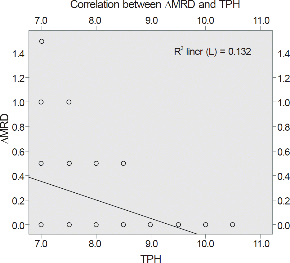 | Figure 4.Significant negative correlationbetween Δ MRD1 and tarsal plate height using correlation analysis (R2=0.132). |
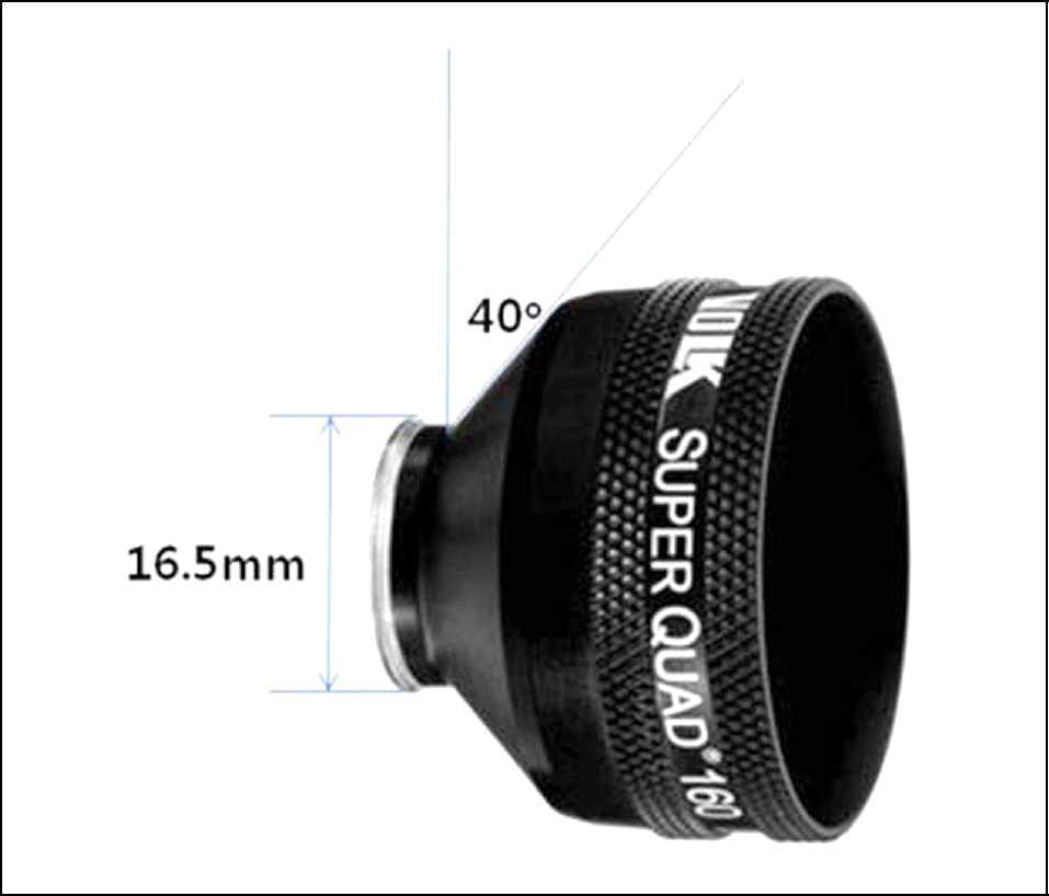 | Figure 5.Superquad 160® contact ophthalmoscopiclens (Volk Optical, Inc., USA). The diameters of the eye contact part of each ophthalmoscopic lens are about 16.5 mm, respectively, and the shaft of each contact ophthalmoscopic lens comes in contact with the upper eyelid at an angle of 40 degrees. |
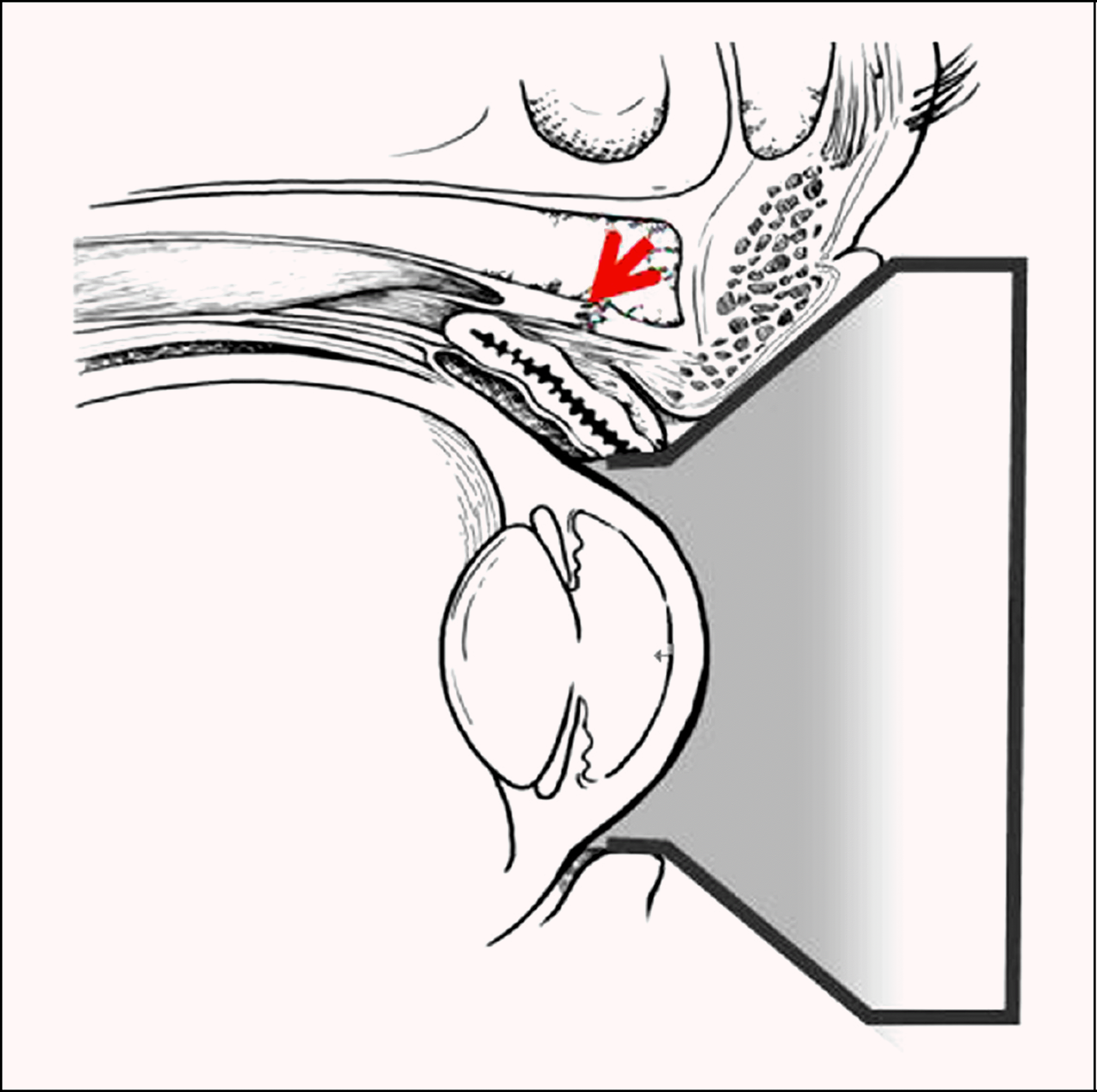 | Figure 6.The illustration shows the relation between the eye-lid and contact ophthalmoscopic lens. When the contact oph-thalmoscopic lens is fitted to the eyes, the lower part of the up-per eyelid is bent against the patient’s eyes and the dehiscence or disinsertion at the insertion part of levatoraponeurosis could occur (arrow). |
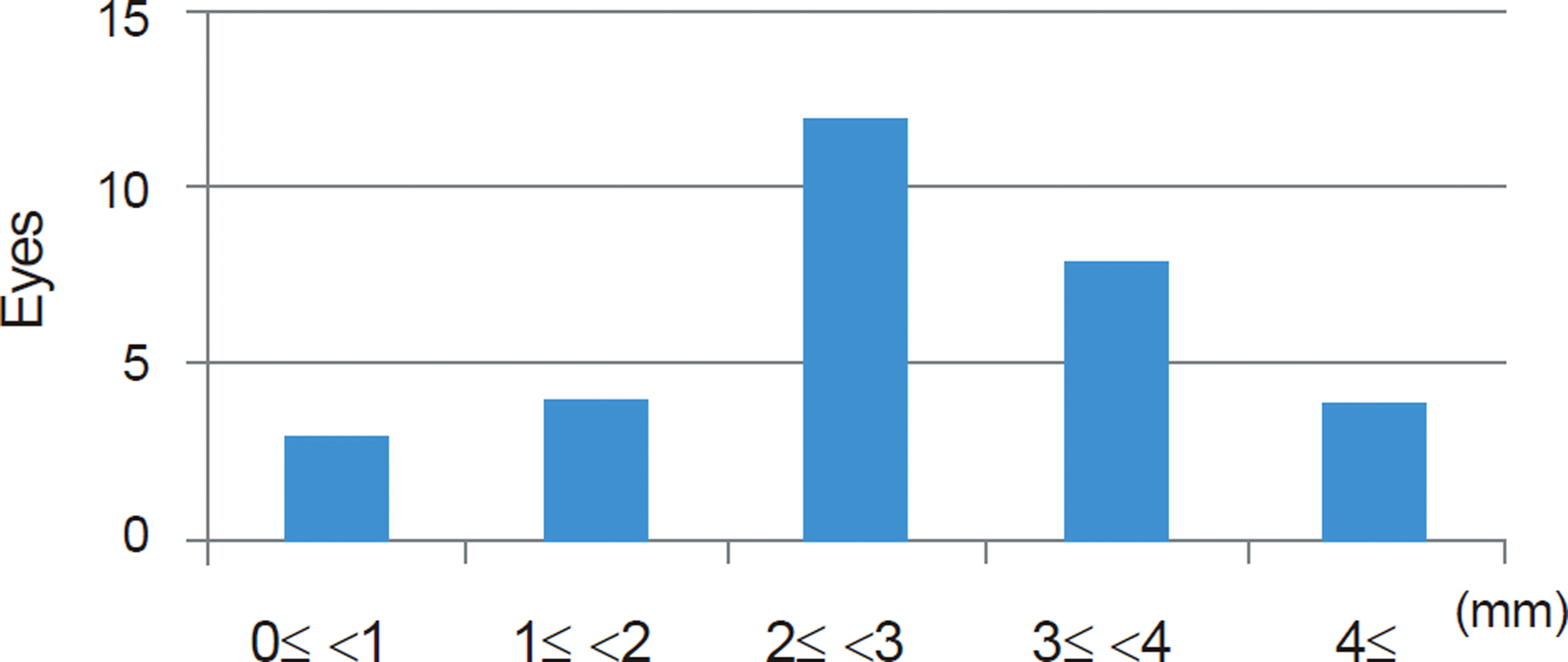
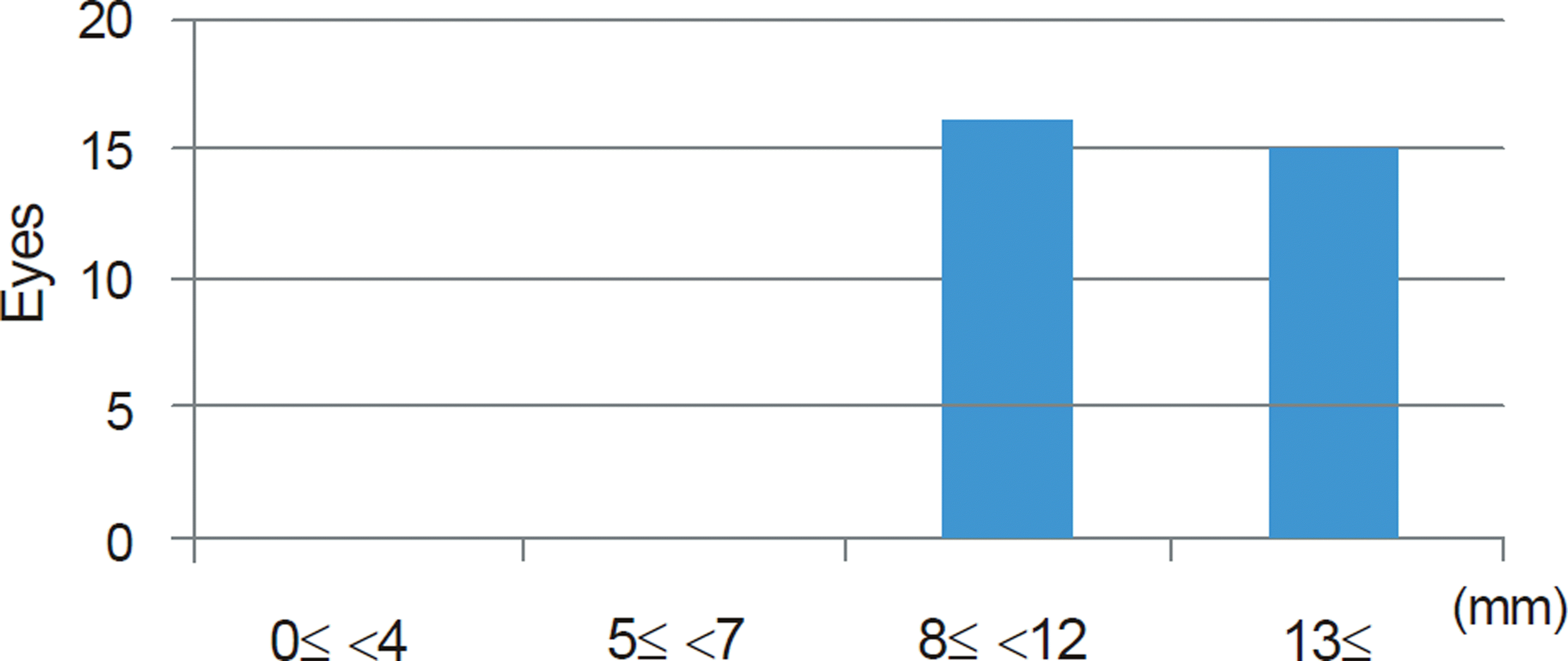
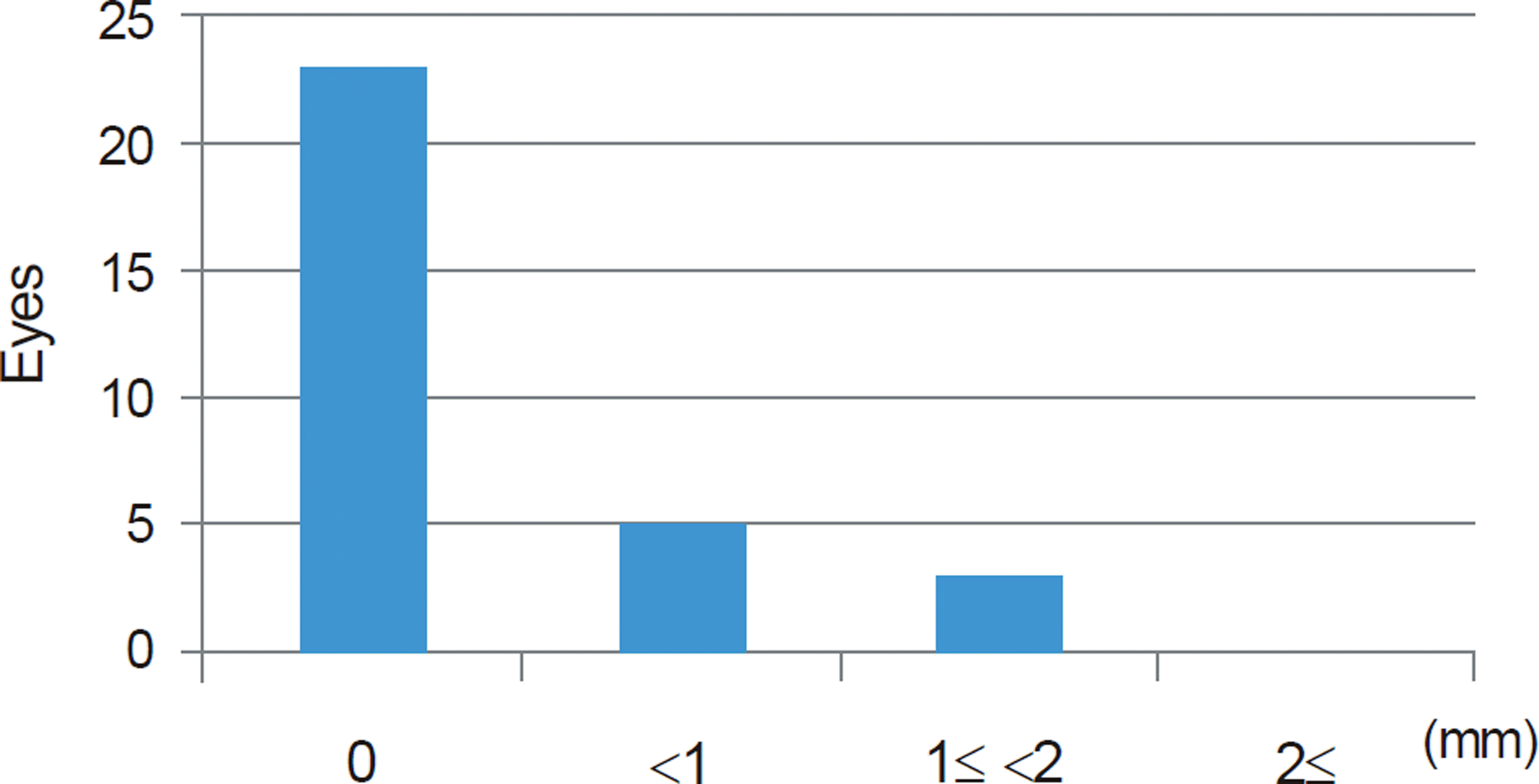
Table 1.
Baseline evaluation of eyelid before panretinal photo-coagulation using ophthalmoscopic contact lens
Table 2.
Changes of MRD1 and levator function, palpebral fissure height after panretinal photocoagulation using ophthalmoscopic contact lens
| PostPRP - PrePRP | p-value* | ||
|---|---|---|---|
| Treated eyes | MRD1 (mm) | -0.2 ± 0.4 (-1.5-0.0) | 0.008 |
| Levator function (mm) | -0.1 ± 0.3 (-2.0-0.0) | 0.063 | |
| Palpebral fissure height (mm) | -0.1 ± 0.3 (-1.0-0.0) | 0.031 | |
| Untreated eyes | MRD1 (mm) | -0.1 ± 0.4 (-1.5-0.5) | 0.203 |
| Levator function (mm) | -0.1 ± 0.2 (-1.5-0.0) | 0.250 | |
| Palpebral fissure height (mm) | -0.1 ± 0.2 (-1.0-0.0) | 0.063 |
 XML Download