

 PDF
PDF ePub
ePub Citation
 Print
Print


Abstract
Purpose
To evaluate the radiographic volume change in extraocular muscles (EOM) following orbital wall decompression for Nunery type 1 and type 2 thyroid-associated orbitopathy (TAO).
Methods
Medical records of 31 orbits in 20 patients undergoing postoperative orbital CT after orbital decompression for TAO were retrospectively reviewed. The patients were divided according to Nunery classifications. A type 1 classification was assigned to patients who had no diplopia and essentially normal versions. A type 2 classification was assigned to patients with restrictive motility loss and diplopia within 20 degrees of the primary position. EOM volumes were determined by the summation of each EOM's cross-sectional areas in the coronal plane of the CT scans and multiplying the sum by the slice thickness. Main outcome measure was a comparison of EOM volume changes between types 1 and 2 TAO and a relationship between EOM volume and change in proptosis.
Results
In type 2 TAO, a significant increase in the volume of the medial rectus muscle, lateral rectus, and total EOM was detected postoperatively (p = 0.044, 0.022, 0.049), whereas no significant changes were found in the EOM's volume changes in type 1 TAO. The reduction of proptosis after orbital decompression in type 1 TAO was significantly greater than in type 2 TAO (p = 0.025). A significant positive association was observed between the preoperative EOM volumes and the reduction of proptosis following orbital wall decompression (r = 0.505).
Go to : 
References
1. Burch HB, Wartofsky L. Graves' ophthalmopathy: current abdominal regarding pathogenesis and management. Endocr Rev. 1993; 14:747–93.
2. Weetman AP. Thyroid-associated eye disease: pathophysiology. Lancet. 1991; 338:25–8.

3. Wiersinga WM, Prummel MF. An evidence-based approach to the treatment of Graves' ophthalmopathy. Endocrinol Metab Clin North Am. 2000; 29:297–319.
4. Bartalena L, Marocci C, Bogazzi F, et al. Glucocorticoid therapy of Graves' ophthalmopathy. Exp Clin Endocrinol. 1991; 97:320–7.
5. Wiersinga WM. Immunosuppressive treatment of Graves' ophthalmopathy. Trends Endocrinol Metab. 1990; 1:377–81.
6. Bartalena L, Pinchera A, Marcocci C. Management of Graves' ophthalmopathy: reality and perspectives. Endocr Rev. 2000; 21:168–99.
7. McCord CD Jr. Current trends in orbital decompression. Ophthalmology. 1985; 92:21–33.
8. Lyons CJ, Rootman J. Orbital decompression for disfiguring exophthalmos in thyroid orbitopathy. Ophthalmology. 1994; 101:223–30.
9. Wenz R, Levine MR, Putterman A, et al. Extraocular muscle abdominal after orbital decompression for Graves' ophthalmopathy. Ophthal Plast Reconstr Surg. 1994; 10:34–41.
10. Hu WD, Annunziata CC, Chokthaweesak W, et al. Radiographic analysis of extraocular muscle volumetric changes in thyroid-abdominal orbitopathy following orbital decompression. Ophthal Plast Reconstr Surg. 2010; 26:1–6.
11. Alsuhaibani AH, Carter KD, Policeni B, Nerad JA. Effect of orbital bony decompression for Graves' orbitopathy on the volume of extraocular muscles. Br J Ophthalmol. 2011; 95:1255–8.
12. Baldeschi L, Lupetti A, Vu P, et al. Reactivation of Graves' abdominal after rehabilitative orbital decompression. Ophthalmology. 2007; 114:1395–402.
13. Kalmann R, Mourits MP. Late recurrence of unilateral graves abdominal on the contralateral side. Am J Ophthalmol. 2002; 133:727–9.
14. Selva D, Chen C, King G. Late reactivation of thyroid orbitopathy. Clin Experiment Ophthalmol. 2004; 32:46–50.
15. Nunery WR. Ophthalmic Graves' disease: a dual theory of pathogenesis. Ophthalmol Clin North Am. 1991; 4:73–87.
16. Nunery WR, Martin RT, Heinz GW, Gavin TJ. The association of cigarette smoking with clinical subtypes of ophthalmic Graves' disease. Ophthal Plast Reconstr Surg. 1993; 9:77–82.
17. Nunery WR, Nunery CW, Martin RT, et al. The risk of diplopia abdominal orbital floor and medial wall decompression in subtypes of ophthalmic Graves' disease. Ophthal Plast Reconstr Surg. 1997; 13:153–60.
18. Park SJ, Kim YD. Two-wall orbital decompression for endocrine exophthalmos. J Korean Ophthalmol Soc. 1992; 33:907–13.
19. Bunting H, Creten O, Muhtaseb M, Shuttleworth G. Late reactivation of thyroid associated ophthalmopathy causing optic neuropathy. Postgrad Med J. 2008; 84:388–90.
20. Chou PI, Feldon SE. Late onset dysthyroid optic neuropathy. Thyroid. 1994; 4:213–6.
21. Dagi LR, Zoumalan CI, Konrad H, et al. Correlation between abdominal muscle size and motility restriction in thyroid eye disease. Ophthal Plast Reconstr Surg. 2011; 27:102–10.
Go to :
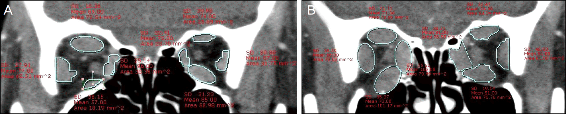 | Figure 1.(A) Orbital CT scan for a patient who had orbital decompression of both orbits. The medial rectus, inferior rectus, lateral rectus, and superior rectus group in the left side was outlined on coronal view. (B) Postoperative coronal CT of same patient documenting postoperative bilateral enlargement in cross-sectional area of medial rectus, inferior rectus, lateral rectus, and superior rectus. |
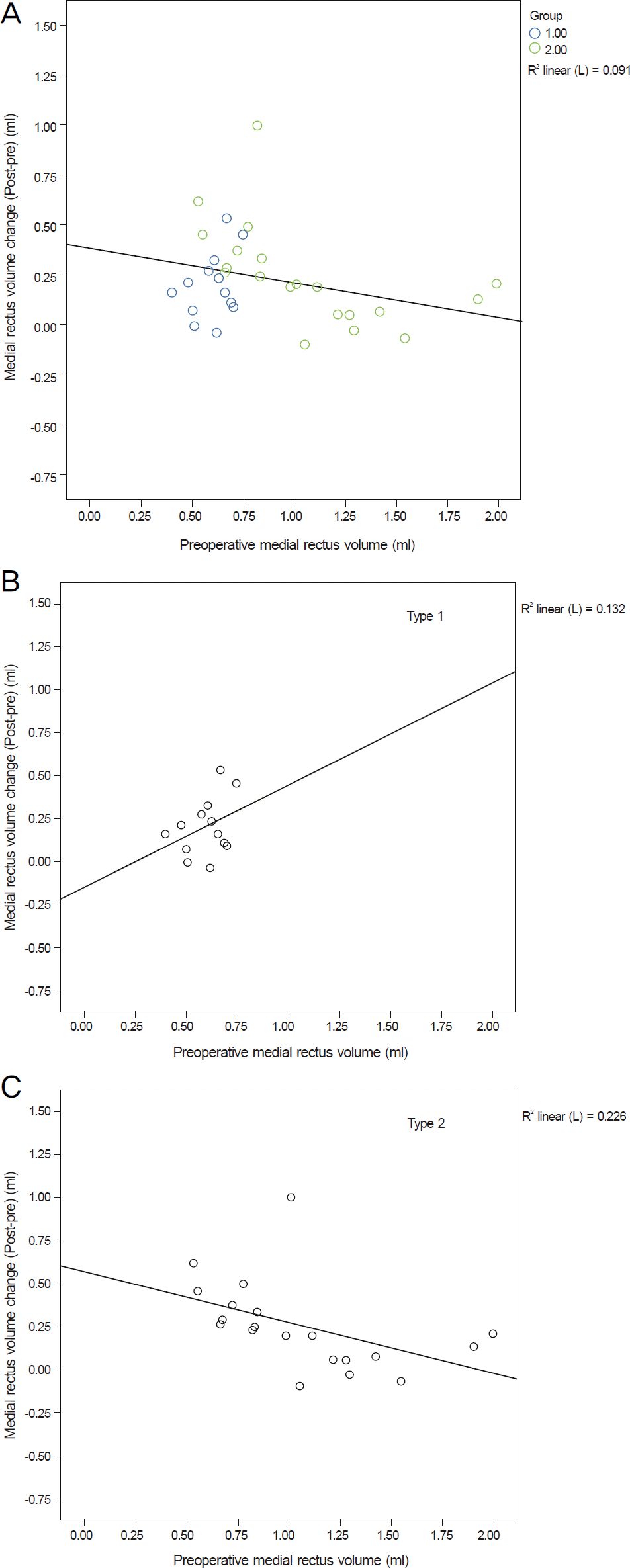 | Figure 2.(A) The relationship between the changes in medial rectus muscle volume (postoperative-preoperative in ml) and the preoperative medial rectus muscle volume in ml. (B) Type 1 C. Type 2. |
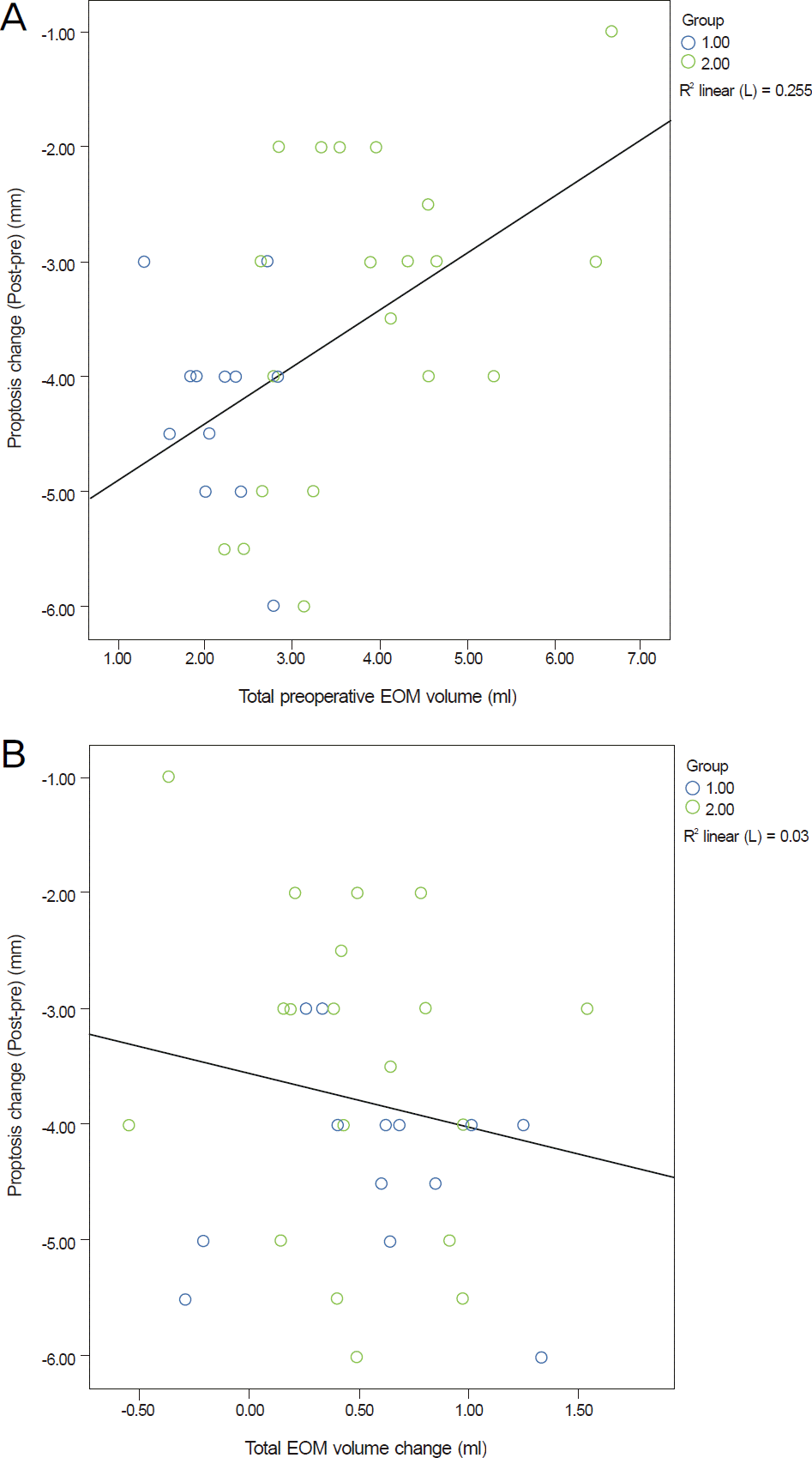 | Figure 3.(A) The relationship between the preoperative total extraocular muscle volume (in ml) and the changes in proptosis (postoperative-preoperative in mm). (B) The relationship between the changes in total extraocular muscle volume (postoperative-preoperative in ml) and the changes in proptosis (postoperative-preoperative in mm). |
Table 1.
Demographic data of Nunery type 1 and type 2 patients
Table 2.
The preoperative and postoperative volume (ml) of the rectus muscles following orbital decompression
| CT measure |
Type 1 (n = 11) |
Type 2 (n = 20) |
||||||
|---|---|---|---|---|---|---|---|---|
| Pre-operative | Post-operative | Change* | p-value | Pre-operative | Post-operative | Change* | p-value | |
| MR | 0.63 ± 0.11 | 0.84 ± 0.4 | 0.21 ± 0.42 | 0.085 | 1.06 ± 0.52 | 1.32 ± 0.49 | 0.26 ± 0.32 | 0.022 |
| LR | 0.48 ± 0.32 | 0.55 ± 0.31 | 0.07 ± 0.17 | 0.225 | 0.71 ± 0.44 | 0.82 ± 0.43 | 0.11 ± 0.13 | 0.049 |
| IR | 0.71 ± 0.17 | 0.77 ± 0.27 | 0.06 ± 0.16 | 0.593 | 1.27 ± 0.44 | 1.30 ± 0.46 | 0.03 ± 0.21 | 0.866 |
| SRG | 0.53 ± 0.16 | 0.57 ± 0.22 | 0.04 ± 0.15 | 0.768 | 0.86 ± 0.43 | 0.88 ± 0.32 | 0.02 ± 0.20 | 0.851 |
| Total EOM† | 2.35 ± 0.58 | 2.73 ± 1.04 | 0.38 ± 0.66 | 0.247 | 3.89 ± 1.51 | 4.32 ± 1.37 | 0.42 ± 0.61 | 0.044 |
 XML Download