

 PDF
PDF ePub
ePub Citation
 Print
Print


Abstract
Purpose
To compare the 1-year outcome of 1.8 mm and 2.2 mm microcoaxial cataract surgery (MCCS) versus 2.75 mm conventional cataract surgery (CCS).
Methods
The present study evaluated 120 eyes (40 eyes in each group). The mean ultrasound power, ultrasound time (UST), and cumulative dissipated energy (CDE) were measured preoperatively and at 1 day, 1, 2, 6 months, and 1 year postoperative. Visual acuity, number of corneal endothelial cells, and surgically induced astigmatism (SIA) were compared.
Results
In LOCS III NO4, 1.8 mm MCCS showed a statistically higher ultrasound time (p-value = 0.031) and CDE (p-value = 0.029), and the day 1 corneal thickness increase was relatively higher in 1.8 mm MCCS (p-value = 0.043) than other two groups. There were no differences in postoperative 1 year visual acuity or number of corneal endothelial cells among the groups. SIA was significantly lower in 1.8 mm and 2.2 mm MCCS compared to that of the conventional treatment (p-value = 0.046).
Conclusions
There were no differences in postoperative 1 year mean endothelial cell density or corrected visual acuity between 1.8 and 2.2 mm MCCS and CCS at all cataract densities. The 1.8 mm and 2.2 mm MCCS techniques were as safe and effective as CCS, and SIA in 1.8 mm and 2.2 mm MCCS was significantly lower than that of CCS.
Figures and Tables
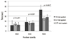 | Figure 1The difference of ultrasound time between 1.8, 2.2 mm MCCS and CCS according to nuclear opacity (LOCS III classification). In LOCS III NO4, 1.8 mm MCCS showed statistically longer ultrasound time compared with other the 2 systems (p-value = 0.031). *Statistically significant.
|
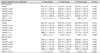
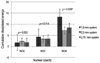 | Figure 2The difference of cumulative dissipitated energy (CDE) between 1.8, 2.2 mm MCCS and CCS according to nuclear opacity. In LOCS III NO4, 1.8 mm MCCS showed statistically higher cumulative dissipated energy compared with the other 2 systems (p-value = 0.029). *Statistically significant.
|
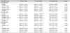
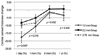 | Figure 3Central corneal thickness change (CCT change) in LOCS III NO4. Postoperative 1 day corneal thickness increased in all groups, and the increase of postoperative 1 day corneal thickness was significantly higher in 1.8 mm MCCS (p-value = 0.043) compared with the other 2 groups. *Statistically significant.
|
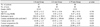
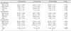
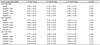
Table 4
Preoperative & postoperative mean endothelial cell density & endothelial cell loss decrement (%) at 1 yr
![]()

References
1. Kelman CD. Phaco-emulsification and aspiration. A new technique of cataract removal. A preliminary report. Am J Ophthalmol. 1967. 64:23–35.
2. Crema AS, Walsh A, Yamane Y, Nosé W. Comparative study of coaxial phacoemulsification and microincision cataract surgery. One-year follow-up. J Cataract Refract Surg. 2007. 33:1014–1018.
3. Ku HC, Kim HJ, Joo CK. The comparison of astigmatism according to the incision size in small incision cataract surgery. J Korean Ophthalmol Soc. 2005. 46:416–421.
4. Jee DH, Lee PY, Joo CK. The comparison of astigmatism according to the incision size in cataract operation. J Korean Ophthalmol Soc. 2003. 44:594–598.
5. Huang FC, Tseng SH. Comparison of surgically induced astigmatism after sutureless temporal clear corneal and scleral frown incisions. J Cataract Refract Surg. 1998. 24:477–481.
6. Lee DS, Joo CK. Effect of incision length on visual recovery and astigmatism in no-suture cataract surgery. J Korean Ophthalmol Soc. 1992. 33:470–475.
7. Hu YJ, Joo CK. Surgically induced astigmatism after temporal clear corneal incision in sutureless cataract surgery. J Korean Ophthalmol Soc. 1998. 39:2622–2627.
8. Vasavada V, Vasavada V, Raj SM, Vasavada AR. Intraoperative performance and postoperative outcomes of microcoaxial phacoemulsification. Observational study. J Cataract Refract Surg. 2007. 33:1019–1024.
9. Osher RH. Microcoaxial phacoemulsification Part 2: clinical study. J Cataract Refract Surg. 2007. 33:408–412.
10. Cavallini GM, Campi L, Masini C, et al. Bimanual microphacoemulsification versus coaxial miniphacoemulsification: prospective study. J Cataract Refract Surg. 2007. 33:387–392.
11. Lee KM, Kwon HG, Joo CK, et al. Microcoaxial cataract surgery outcomes: Comparison of 1.8 mm system and 2.2 mm system. J Cataract Refract Surg. 2009. 35:874–880.
12. Dosso AA, Cottet L, Burgener ND, Di Nardo S. Outcomes of coaxial microincision cataract surgery versus conventional coaxial cataract surgery. J Cataract Refract Surg. 2008. 34:284–288.
13. Alió J, Rodríguez-Prats JL, Galal A, Ramzy M. Outcomes of microincision cataract surgery versus coaxial phacoemulsification. Ophthalmology. 2005. 112:1997–2003.
14. Kim HJ, Kim JH, Lee DH. Endothelial cell damage in microincision cataract surgery and coaxial phacoemulsification. J Korean Ophthalmol Soc. 2007. 48:19–26.
15. Can I, Takmaz T, Yildiz Y, et al. Coaxial, microcoaxial, and biaxial microincision cataract surgery: prospective comparative study. J Cataract Refract Surg. 2010. 36:740–746.
16. Choi JA, Chung SK, Kim HS. Comparative study of microcoaxial cataract surgery and conventional cataract surgery. J Korean Ophthalmol Soc. 2008. 49:904–910.
17. Cravy TV. Calculation of the change in corneal astigmatism following cataract extraction. Ophthalmic Surg. 1979. 10:38–49.
18. Kurz S, Krummenauer F, Gabriel P, et al. Biaxial microincision versus coaxial small-incision clear cornea cataract surgery. Ophthalmology. 2006. 113:1818–1826.
19. Lundberg B, Jonsson M, Behndig A. Postoperative corneal swelling correlates strongly to corneal endothelial cell loss after phacoemulsification cataract surgery. Am J Ophthalmol. 2005. 139:1035–1041.
20. Hayashi K, Hayashi H, Nakao F, Hayashi F. Risk factors for corneal endothelial injury during phacoemulsification. J Cataract Refract Surg. 1996. 22:1079–1084.
21. Dick HB, Kohnen T, Jacobi FK, Jacobi KW. Long-term endothelial cell loss following phacoemulsification through a temporal clear corneal incision. J Cataract Refract Surg. 1996. 22:63–71.
22. Joussen AM, Barth U, Cubuk H, Koch H. Effect of irrigating solution and irrigation temperature on the cornea and pupil during phacoemulsification. J Cataract Refract Surg. 2000. 26:392–397.
23. Bourne RR, Minassian DC, Dart JK, et al. Effect of cataract surgery on the corneal endothelium: modern phacoemulsification compared with extracapsular cataract surgery. Ophthalmology. 2004. 111:679–685.
24. Suzuki H, Takahashi H, Hori J, et al. Phacoemulsification associated corneal damage evaluated by corneal volume. Am J Ophthalmol. 2006. 142:525–528.
25. Yao K, Tang X, Ye P. Corneal astigmatism, high order aberrations, and optical quality after cataract surgery: microincision versus small incision. J Refract Surg. 2006. 22:9 Suppl. S1079–S1082.
 XML Download