

 PDF
PDF ePub
ePub Citation
 Print
Print


Abstract
Purpose
To investigate mean keratometric and refractive value after penetrating keratoplasty according to the difference between donor and recipient cornea size.
Methods
In a retrospective study, Keratoconus patients who underwent penetrating keratoplasty for keratoconus from January 2005 to July 2008 were examined. Preoperatively, axial length was measured using applanation ultrasonography and anterior chamber depth, white to white diameter were also measured using the corneal topography. The trephine size of donor and recipient during the surgery were recorded. Preoperatively, 6, 12, and 24 months postoperatively refraction and keratometric value were evaluated in groups divided according to corneal trephine size difference.
Results
Among the 41 eyes of 41 patients, there was a mean age at transplant of 26.4 years. After surgery, the decrease of anterior chamber depth could contribute to the decrease of myopic change. However, the differences between donor and recipient cornea size do not have a significant affect on postoperative keratometric or refractive value.
Figures and Tables
Figure 1
The keratometric value changes of groups divided according to the trephine size difference at 6, 12 and 24 months postoperatively after conventional penetrating keratoplasty. There was no significant keratometric value difference among these groups at postoperative 6 months, 12 months and 24 months.
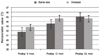
Figure 2
The spherical equivalent changes of groups divided according to the trephine size difference at 6, 12 and 24 months postoperatively after conventional penetrating keratoplasty. There was no significant refractive value difference among these groups at postoperative 6 months, 12 months and 24 months.
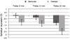
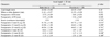
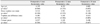
Table 2
Postoperative data of patients according to the axial length and corneal trephine size difference

References
1. Lee KM, Chung SK. Studies of patients maintaining clear cornea over 20 years following penetrating keratoplasty. J Korean Ophthalmol Soc. 2009. 50:19–26.
2. Sarhan AR, Dua HS, Beach M. Effect of disagreement between refractive, keratometric, and topographic determination of astigmatic axis on suture removal after penetrating keratoplasty. Br J Ophthalmol. 2000. 84:837–841.
3. Perry HD, Foulks GN. Oversize donor buttons in corneal transplantation surgery for keratoconus. Ophthalmic Surg. 1987. 18:751–752.
4. Duran JA, Malvar A, Diez E. Corneal dioptric power after penetrating keratoplasty. Br J Ophthalmol. 1989. 73:657–660.
5. Wilson SE, Bourne WM. Effect of recipient-donor trephine size disparity on refractive error in keratoconus. Ophthalmology. 1989. 96:299–305.
6. Lanier JD, Bullington RH Jr, Prager TC. Axial length in keratoconus. Cornea. 1992. 11:250–254.
7. Javadi MA, Mohammadi MJ, Mirdehghan SA, Sajjadi SH. A comparison between donor-recipient corneal size and its effect on the ultimate refractive error induced in keratoconus. Cornea. 1993. 12:401–405.
8. Goble RR, Hardman Lea SJ, Falcon MG. The use of the same size host and donor trephine in penetrating keratoplasty for keratoconus. Eye (Lond). 1994. 8(Pt 3):311–314.
9. Tuft SJ, Fitzke FW, Buckley RJ. Myopia following penetrating keratoplasty for keratoconus. Br J Ophthalmol. 1992. 76:642–645.
10. Lim L, Pesudovs K, Coster DJ. Penetrating keratoplasty for keratoconus: visual outcome and success. Ophthalmology. 2000. 107:1125–1131.
11. Yoon JW, Chung SK, Rhee SW. Clinical results of penetrating keratoplasty in keratoconus. J Korean Ophthalmol Soc. 1993. 34:85–90.
12. Son JH, Tchah H, Kim YJ. Suture tension adjustment of single running suture in penetrating keratoplasty. J Korean Ophthalmol Soc. 1993. 34:198–201.
13. Jeong HS, Kim WJ, Lee JH. The comparison of long term clinical effect between penetrating keratoplasty and epikeratoplasty on keratoconus. J Korean Ophthalmol Soc. 1994. 35:646–651.
14. Kim KE, Joo CK. Changes in astigmatism after suture removal in penetrating keratoplasty. J Korean Ophthalmol Soc. 2003. 44:284–288.
15. Han ES, Wee WR, Lee JH, Kim MK. Changes in corneal curvature after suture removal in penetrating keratoplasty. J Korean Ophthalmol Soc. 2006. 47:1543–1548.
16. Kil SJ, Choi SH. The change of keratometric value following penetrating keratoplasty. J Korean Ophthalmol Soc. 1998. 39:1984–1990.
17. Rabinowitz YS, McDonnell PJ. Computer-assisted corneal topography in keratoconus. Refract Corneal Surg. 1989. 5:400–408.
18. Troutman RC, Meltzer M. Astigmatism and myopia in keratoconus. Trans Am Ophthalmol Soc. 1972. 70:265–277.
19. Jensen AD, Maumenee AE. Refractive errors following keratoplasty. Trans Am Ophthalmol Soc. 1974. 72:123–131.
20. Perl T, Charlton KH, Binder PS. Disparate diameter grafting. Astigmatism, intraocular pressure, and visual acuity. Ophthalmology. 1981. 88:774–781.
21. Girard LJ, Eguez I, Esnaola N, et al. Effect of penetrating keratoplasty using grafts of various sizes on keratoconic myopia and astigmatism. J Cataract Refract Surg. 1988. 14:541–547.
 XML Download