

 PDF
PDF ePub
ePub Citation
 Print
Print


Abstract
Purpose
To report 2 cases of far-advanced keratoconus with a high value of maximum keratometry and very thin corneas treated with corneal crosslinking (CXL).
Case summary
The thinnest corneal thickness of an 18-year-old woman with maximum keratometry of 106.5 D (case 1) was 335 µm. The thinnest corneal thickness of a 43-year-old man with maximum keratometry of 120.3 D (case 2) was 345 µm. The two cases underwent a customized topography and pachymetry-guided epithelial debridement technique to preserve the epithelium where the cornea was within 2 mm around the cone and subsequent CXL. Postoperative maximum keratometry was 97.2 D 24 months after CXL in case 1 and 109.3 D 18 months after CXL in case 2. Postoperatively, the thinnest corneal thickness was 343 µm in case 1 and 162 µm in case 2. The corneal thickness in case 1 was stabilized during the follow-up examination. The pupil center and apex of the corneal thickness in case 2 with the higher maximum keratometry was stabilized, but the thinnest corneal thickness was decreased immediately after CXL and did not recover before CXL.
Conclusions
CXL was performed in 2 cases of far-advanced keratoconus. Results showed reduced maximum keratometry but, variable values in corneal thickness during the follow-up examination in the 2 cases. Longer follow-up is necessary, and CXL should be performed cautiously, especially for patients with far-advanced keratoconus.
Figures and Tables
Figure 1
Anterior segment photography of the left eye in case 1 showing the apex of the cone (white arrow) in the inferior paracentral cornea and anterior stromal scars (white arrow head) within the cone at initial visit.
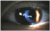
Figure 2
Corneal topography of the left cornea in case 1. (A) shows the central cornea with high maximum K (Kmax) values of 106.5 D and mean K (Kmean) of 74.1 D at initial visit. (B) shows Kmax of 97.2 D and Kmean of 72.2 D 24 months after CXL. (C) shows corneal thickness at the pupil center (CCT) of 347 µm, corneal thickness at the apex (CTapex) of 351 µm and corneal thickness at the thinnest point (CTthin) of 335 µm. The outer circle was placed at the 9 mm corneal zone and the area of the cornea within the inner gray line was placed under 400 µm of corneal thickness at initial visit. (D) shows CCT of 387 µm, CTapex of 361 µm, and CTthin of 343 µm 24 months after CXL.
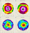
Figure 3
Anterior segment photography of the left eye in case 2 showing the apex of the cone (white arrow) in the inferior paracentral cornea and anterior stromal scars (white arrow head) lateral to the cone at initial visit.

Figure 4
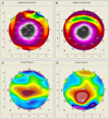
Corneal topography of the left cornea in case 2. (A) shows Kmax of 120.3 D and Kmean of 82.7 D at initial visit. (B) shows Kmax of 109.3 D and Kmean of 80.1 D 18 months after CXL. (C) shows CCT of 459 µm, CTapex of 403 µm, and CTthin of 345 µm, the outer circle was placed at the 9 mm corneal zone and the area of cornea within the inner gray line was placed under 400 µm of corneal thickness at initial visit. (D) shows CCT of 452 µm, CTapex of 403 µm, and CTthin of 162 µm 18 months after CXL.
References
1. Krachmer JH, Feder RS, Belin MW. Keratoconus and related noninflammatory corneal thinning disorders. Surv Ophthalmol. 1984. 28:293–322.
2. Spoerl E, Huhle M, Seiler T. Induction of cross-links in corneal tissue. Exp Eye Res. 1998. 66:97–103.
3. Wollensak G, Spoerl E, Seiler T. Stress-strain measurements of human and porcine corneas after riboflavin-ultraviolet-A-induced cross-linking. J Cataract Refract Surg. 2003. 29:1780–1785.
4. Spoerl E, Wollensak G, Seiler T. Increased resistance of crosslinked cornea against enzymatic digestion. Curr Eye Res. 2004. 29:35–40.
5. Spoerl E, Mrochen M, Sliney D, et al. Safety of UVA-riboflavin cross-linking of the cornea. Cornea. 2007. 26:385–389.
6. Alió JL, Shabayek MH. Corneal higher order aberrations: a method to grade keratoconus. J Refract Surg. 2006. 22:539–545.
7. Kymionis GD, Diakonis VF, Coskunseven E, et al. Customized pachymetric guided epithelial debridement for corneal collagen cross linking. BMC Ophthalmol. 2009. 9:10.
8. Nottingham J. Practical Observations on Conical Cornea: and on the Short Sight, and Other Defects of Vision Connected with It. 1854. London: J Churchill.
9. Romero-Jiménez M, Santodomingo-Rubido J, Wolffsohn JS. Keratoconus: a review. Cont Lens Anterior Eye. 2010. 33:157–166.
10. Tomkins O, Garzozi HJ. Collagen cross-linking: strengthening the unstable cornea. Clin Ophthalmol. 2008. 2:863–867.
11. Vinciguerra P, Albè E, Trazza S, et al. Refractive, topographic, tomographic, and aberrometric analysis of keratoconic eyes undergoing corneal cross-linking. Ophthalmology. 2009. 116:369–378.
12. Grewal DS, Brar GS, Jain R, et al. Corneal collagen crosslinking using riboflavin and ultraviolet-A light for keratoconus: one-year analysis using Scheimpflug imaging. J Cataract Refract Surg. 2009. 35:425–432.
13. Raiskup F, Hoyer A, Spoerl E. Permanent corneal haze after riboflavin-UVA-induced cross-linking in keratoconus. J Refract Surg. 2009. 25:S824–S828.
14. Koller T, Mrochen M, Seiler T. Complication and failure rates after corneal crosslinking. J Cataract Refract Surg. 2009. 35:1358–1362.
15. Hafezi F, Mrochen M, Iseli HP, Seiler T. Collagen crosslinking with ultraviolet-A and hypoosmolar riboflavin solution in thin corneas. J Cataract Refract Surg. 2009. 35:621–624.
16. Raiskup F, Spoerl E. Corneal cross-linking with hypo-osmolar riboflavin solution in thin keratoconic corneas. Am J Ophthalmol. 2011. 152:28–32.
17. Wollensak G, Iomdina E. Biomechanical and histological changes after corneal crosslinking with and without epithelial debridement. J Cataract Refract Surg. 2009. 35:540–546.
18. Tu KL, Aslanides IM. Orbscan II anterior elevation changes following corneal collagen cross-linking treatment for keratoconus. J Refract Surg. 2009. 25:715–722.
19. Greenstein SA, Shah VP, Fry KL, Hersh PS. Corneal thickness changes after corneal collagen crosslinking for keratoconus and corneal ectasia: one-year results. J Cataract Refract Surg. 2011. 37:691–700.
20. Koller T, Iseli HP, Hafezi F, et al. Scheimflug imaging of corneas after collagen cross-linking. Cornea. 2009. 28:510–515.
21. Bottós KM, Dreyfuss JL, Regatieri CV, et al. Immunofluorescence confocal microscopy of porcine corneas following collagen cross-linking treatment with riboflavin and ultraviolet A. J Refract Surg. 2008. 24:S715–S719.
22. Mazzotta C, Traversi C, Baiocchi S, et al. Conservative treatment of keratoconus by riboflavin-uva-induced cross-linking of corneal collagen: qualitative investigation. Eur J Ophthalmol. 2006. 16:530–535.
23. Caporossi A, Baiocchi S, Mazzotta C, et al. Parasurgical therapy for keratoconus by riboflavin-ultraviolet type A rays induced cross-linking of corneal collagen: preliminary refractive results in an Italian study. J Cataract Refract Surg. 2006. 32:837–845.
24. Shi W, Li S, Gao H, et al. Modified deep lamellar keratoplasty for the treatment of advanced-stage keratoconus with steep curvature. Ophthalmology. 2010. 117:226–231.
25. Sutton G, Hodge C, McGhee CN. Rapid visual recovery after penetrating keratoplasty for keratoconus. Clin Experiment Ophthalmol. 2008. 36:725–730.
26. Inj JJ, Ing HH, Nelson LR, et al. Ten-year postoperative results of penetrating keratoplasty. Ophthalmology. 1998. 105:1855–1865.
27. Kim KH, Ahn K, Chung ES, et al. Comparison of deep anterior lamellar keratoplasty and penetrating keratoplasty for keratoconus. J Korean Ophthalmol Soc. 2008. 49:222–229.
 XML Download