

 PDF
PDF ePub
ePub Citation
 Print
Print


Abstract
Purpose
To evaluate the relations between progression of glaucoma in visual field and peripapillary area change in normal tension glaucoma (NTG).
Methods
We respectively evaluated 66 patients (66 eyes) with normal tension glaucoma; these patients were classified as progressive glaucoma group and non-progressive glaucoma group by visual field test. Interobserver and intraobserver agreement was evaluated for qualitative assessment. Zone β change was measured by Image J (National Institute of Health, Bethesda, USA) by two methods. One method used paired t-test and the other method used CV (correlation of variation) to define significant progression of zone β.
Figures and Tables
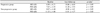
Figure 1
Scatterplots showing the interobserver agreement between the two observers in measuring areas (pixel) of progression of peripapillary atrophy (beta zone).
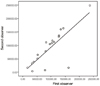
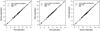
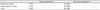
Figure 2
Scatterplots showing the intraobserver agreement between each observation in measuring areas (log pixel) of progression of peripapillary atrophy (beta zone).

References
1. Armaly MF. Ocular pressure and visual fields. A ten-year follow-up study. Arch Ophthalmol. 1969. 81:25–40.
2. Levene RZ. Low tension glaucoma: a critical review and new material. Surv Ophthalmol. 1980. 24:621–664.
3. Perkins ES. The Bedford glaucoma survey. I. Long-term follow-up of borderline cases. Br J Ophthalmol. 1973. 57:179–185.
4. Van Buskirk EM, Cioffi GA. Glaucomatous optic neuropathy. Am J Ophthalmol. 1992. 113:447–452.
5. Brubaker RF. Delayed functional loss in glaucoma. LII Edward Jackson Memorial Lecture. Am J Ophthalmol. 1996. 121:473–483.
6. Araie M, Sekine M, Suzuki Y, Koseki N. Factors contributing to the progression of visual field damage in eyes with normal-tension glaucoma. Ophthalmology. 1994. 101:1440–1444.
7. Collaborative Normal-Tension Glaucoma Study Group. Comparison of glaucomatous progression between untreated patients with normal-tension glaucoma and patients with therapeutically reduced intraocular pressures. Am J Ophthalmol. 1998. 126:487–497.
8. Crichton A, Drance SM, Douglas GR, Schulzer M. Unequal intraocular pressure and its relation to asymmetric visual field defects in low-tension glaucoma. Ophthalmology. 1989. 96:1312–1314.
9. Daugeliene L, Yamamoto T, Kitazawa Y. Risk factors for visual field damage progression in normal-tension glaucoma eyes. Graefes Arch Clin Exp Ophthalmol. 1999. 237:105–108.
10. Nakagami T, Yamazaki Y, Hayamizu F. Prognostic factors for progression of visual field damage in patients with normal-tension glaucoma. Jpn J Ophthalmol. 2006. 50:38–43.
11. Meyer JH, Brandi-Dohrn J, Funk J. Twenty four hour blood pressure monitoring in normal tension glaucoma. Br J Ophthalmol. 1996. 80:864–867.
12. Jonas JB, Nguyen XN, Gusek GC, Naumann GO. Parapapillary chorioretinal atrophy in normal and glaucoma eyes. I. Morphometric data. Invest Ophthalmol Vis Sci. 1989. 30:908–918.
13. Jonas JB, Fernández MC, Naumann GO. Glaucomatous parapapillary atrophy. Occurrence and correlations. Arch Ophthalmol. 1992. 110:214–222.
14. Jonas JB, Naumann GO. Parapapillary chorioretinal atrophy in normal and glaucoma eyes. II. Correlations. Invest Ophthalmol Vis Sci. 1989. 30:919–926.
15. Park KH, Tomita G, Liou SY, Kitazawa Y. Correlation between peripapillary atrophy and optic nerve damage in normal-tension glaucoma. Ophthalmology. 1996. 103:1899–1906.
16. Tezel G, Kass MA, Kolker AE, Wax MB. Comparative optic disc analysis in normal pressure glaucoma, primary open-angle glaucoma, and ocular hypertension. Ophthalmology. 1996. 103:2105–2113.
17. Kono Y, Zangwill L, Sample PA, et al. Relationship between parapapillary atrophy and visual field abnormality in primary open-angle glaucoma. Am J Ophthalmol. 1999. 127:674–680.
18. Primrose J. Early signs of the glaucomatous disc. Br J Ophthalmol. 1971. 55:820–825.
19. Rockwood EJ, Anderson DR. Acquired peripapillary changes and progression in glaucoma. Graefes Arch Clin Exp Ophthalmol. 1988. 226:510–515.
20. Tuulonen A, Jonas JB, Välimäki S, et al. Interobserver variation in the measurements of peripapillary atrophy in glaucoma. Ophthalmology. 1996. 103:535–541.
21. See JL, Nicolela MT, Chauhan BC. Rates of neuroretinal rim and peripapillary atrophy area change: a comparative study of glaucoma patients and normal controls. Ophthalmology. 2009. 116:840–847.
22. Hodapp E, Parrish RK, Anderson DR. Clinical Decisions in Glaucoma. 1993. St. Louis: Mosby;52–61.
23. Uchida H, Ugurlu S, Caprioli J. Increasing peripapillary atrophy is associated with progressive glaucoma. Ophthalmology. 1998. 105:1541–1545.
24. Park KH, Park SJ, Lee YJ, et al. Ability of peripapillary atrophy parameters to differentiate normal-tension glaucoma from glaucomalike disk. J Glaucoma. 2001. 10:95–101.
25. Kono Y, Jonas JB, Zangwill L, et al. Agreement of measurement of parapapillary atrophy with confocal scanning laser ophthalmoscopy and planimetry of photographs. J Glaucoma. 1999. 8:105–110.
26. Jonas JB. Clinical implications of peripapillary atrophy in glaucoma. Curr Opin Ophthalmol. 2005. 16:84–88.
27. Hayreh SS, Jonas JB, Zimmerman MB. Parapapillary chorioretinal atrophy in chronic high-pressure experimental glaucoma in rhesus monkeys. Invest Ophthalmol Vis Sci. 1998. 39:2296–2303.
28. Airaksinen PJ, Juvala PA, Tuulonen A, et al. Krieglstein GK, editor. Change of peripapillary atrophy in glaucoma. Glaucoma Update III. 1987. Berlin; New York: Springer-Verlag;97–102.
 XML Download