

 PDF
PDF ePub
ePub Citation
 Print
Print


Abstract
Purpose
The authors of the present study describe a rare case of angiolymphoid hyperplasia with eosinophilia (ALHE) of the eyelid.
Case summary
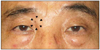
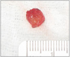
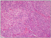
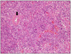
A 63-year-old male who was diagnosed with ALHE based on biopsy of an inguinal mass presented with an eyelid mass of 1 month duration. A light brown, solitary, 1.0 × 0.5 cm-sized mass involved the right upper eyelid. There was no lymphadenopathy, but eosinophilia was present. An excisional biopsy of the mass was performed for diagnosis and management. Macroscopic examination of the excised mass revealed a well-defined, smooth, firm, yellowish-red colored lesion measuring 1.0 × 0.6 × 0.5 cm. Histopathology showed the proliferation of small blood vessels, many of which were lined by enlarged endothelial cells with uniform ovoid nuclei and intracytoplasmic vacuoles. The distinctive endothelial cells were described as having a cobblestone appearance. In addition, a perivascular and interstitial infiltrate composed primarily of lymphocytes and eosinophils was present. ALHE was finally confirmed with clinical and microscopic examination.
Figures and Tables
Figure 1
A relaltively well-demarcated, erythematous, firm, subcutaneous nodule in the upper eyelid.
References
1. Wells GC, Whimster IW. Subcutaneous angiolymphoid hyperplasia with eosinophilia. Br J Dermatol. 1969. 81:1–14.
2. Kim SM, Yoon J, Yoon TJ. Angiolymphoid hyperplasia with eosinophilia on the palm. Ann Dermatol. 2010. 22:358–361.
3. Olsen TG, Helwig EB. Angiolymphoid hyperplasia with eosinophilia. A clinicopathologic study of 116 patients. J Am Acad Dermatol. 1985. 12(5 Pt 1):781–796.
4. Park Y, Chung J, Cho CG. Angiolymphoid hyperplasia with eosinophilia of the tongue: report of a case and review of the literature. Oral Oncol. 2002. 38:103–106.
5. Park JS, Lee MJ. A case of angiolymphoid hyperplasia with eosinophilia (ALHE) in the genital area accompanied by varicocele. Int J Dermatol. 2009. 48:1264–1266.
6. Lee WJ, Kim MS, Lee MW, et al. Angiolymphoid hyperplasia with eosinophilia associated with arteriovenous malformation. Clin Exp Dermatol. 2009. 34:e272–e273.
7. Kim SM, Yoon J, Yoon TJ. Angiolymphoid hyperplasia with eosinophilia on the Palm. Ann Dermatol. 2010. 22:358–361.
8. Jang KA, Lee JY, Kim CH, et al. Angiolymphoid hyperplasia with eosinophilia and Kimura's disease: a clinico-pathologic study in Korea. Korean J Dermatol. 2001. 39:309–317.
9. Thompson MJ, Whitehead J, Gunkel JL, Kulkarni AD. Angiolymphoid hyperplasia with eosinophilia affecting the eyelids. Arch Ophthalmol. 2007. 125:987.
10. Lin B, Tan SH, Looi A. Angiolymphoid hyperplasia with eosinophilia of eyelid with spontaneous regression. Ophthal Plast Reconstr Surg. 2008. 24:308–310.
11. Mariatos G, Gorgoulis VG, Laskaris G, Kittas C. Epithelioid haemangioma (angiolymphoid hyperplasia with eosinophilia) in the inner canthus. J Eur Acad Dermatol Venereol. 2001. 15:90–91.
12. Jang KA, Lee JY, Kim CH, et al. Angiolymphoid hyperplasia with eosinophilia and Kimura's disease: a clinico-pathologic study in Korea. Korean J Dermatol. 2001. 39:309–317.
13. Kitamura H, Ito S, Kuwana N, Yutani C. Epithelioid hemangioma of the temporal artery clinically mimicking temporal arteritis. Pathol Int. 1999. 49:831–835.
14. Rosai J, Gold J, Landy R. The histiocytoid hemangiomas. A unifying concept embracing several previously described entities of skin, soft tissue, large vessels, bone, and heart. Hum Pathol. 1979. 10:707–730.
15. Rosai J. Angiolymphoid hyperplasia with eosinophilia of the skin. Its nosological position in the spectrum of histiocytoid hemangioma. Am J Dermatopathol. 1982. 4:175–184.
16. Weiss SW, Enzinger FM. Epithelioid hemangioendothelioma: a vascular tumor often mistaken for a carcinoma. Cancer. 1982. 50:970–981.
17. Chung DH, Kim BJ, Kim YD. Kimuras disease involving the eyelid and orbit. J Korean Ophthalmol Soc. 2002. 43:1789–1796.
18. Urabe A, Tsuneyoshi M, Enjoji M. Epithelioid hemangioma versus Kimuras disease. A comparative clinicopathologic study. Am J Surg Pathol. 1987. 11:758–766.
19. Googe PB, Harris NL, Mihm MC Jr. Kimura's disease and angiolymphoid hyperplasia with eosinophilia: two distinct histopathological entities. J Cutan Pathol. 1987. 14:263–271.
20. Googe PB, Harris NL, Mihm MC Jr. Kimura's disease and angiolymphoid hyperplasia with eosinophilia: two distinct histopathological entities. J Cutan Pathol. 1987. 14:263–271.
21. Kimura T, Yoshimura S, Ishikawa E. Unusual granulation combined with hyperplastic change of lymphoid tissues. Trans Soc Pathol Jpn. 1948. 37:179–180.
22. Kung IT, Gibson JB, Bannatyne PM. Kimura's disease: a clinico-pathological study of 21 cases and its distinction from angiolymphoid hyperplasia with eosinophilia. Pathology. 1984. 16:39–44.
23. Takenaka T, Okuda M, Usami A, et al. Histological and immunological studies on eosinophilic granuloma of soft tissue, so-called Kimura's disease. Clin Allergy. 1976. 6:27–39.
24. Baghestani S, Firooz A, Ghazisaidi MR. A refractory case of angiolymphoid hyperplasia with eosinophilia successfully treated by surgery. J Dermatolog Treat. 2011. 22:49–51.
25. Satpathy A, Moss C, Raafat F, Slator R. Spontaneous regression of a rare tumour in a child: angiolymphoid hyperplasia with eosinophilia of the hand: case report and review of the literature. Br J Plast Surg. 2005. 58:865–868.
26. Lin B, Tan SH, Looi A. Angiolymphoid hyperplasia with eosinophilia of the eyelid with spontaneous regression. Ophthal Plast Reconstr Surg. 2008. 24:308–310.
27. Archer KF, Hurwitz JJ, Heathcote G. Orbital angiolymphoid hyperplasia with eosinophilia. Presentation as chalazion. Ophthal Plast Reconstr Surg. 1991. 7:208–221.
28. Ruckenstein MJ, Birt BD, Gruss JS. Angiolymphoid hyperplasia with eosinophilia: a case report and literature review. J Otolaryngol. 1989. 18:236–240.
 XML Download