

 PDF
PDF ePub
ePub Citation
 Print
Print


Abstract
Purpose
To compare the ability of three dimensional spectral-domain optical coherence tomography (3D OCT) and Stratus OCT to detect early glaucoma.
Methods
The optic disc topographic and retinal nerve fiber layer (RNFL) thickness parameters were measured by 3D OCT and Stratus OCT in 69 normal eyes and 48 early glaucoma eyes. The discriminating abilities of the two techniques for detection of glaucoma were compared by the area under the receiver operating characteristic curves (AUC).
Results
The best Stratus OCT parameters and criterion that differentiated normal from early glaucoma based on AUC were horizontal integrated rim width (0.85) for optic nerve head parameters, inferior quadrant (0.88) for RNFL parameters, and ≥1 clock-hour abnormal at the 5% level (0.81) based on the normative database for criteria. The best 3D OCT parameters and criterion that differentiated normal from early glaucoma were vertical cup-to-disc ratio (0.85), 11 o'clock RNFL thickness (0.86), and ≥1 clock-hour abnormal at the 1% level (0.78), respectively. When all corresponding the best parameters and criterion were compared, there were no significant differences between the AUCs for Stratus OCT and 3D OCT (p = 0.95, p = 0.73, p = 0.45, respectively).
Figures and Tables
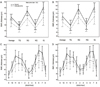 | Figure 1Comparison of Stratus OCT (A, C) and 3D OCT (B, D) in measurement of peripapillary retinal nerve fiber layer (RNFL) thickness on average, in each of 4 quadrants (A, B), in each of the 12 clock-hour sectors (C, D) between normal group and early glaucoma group. TQ = temporal quadrant; SQ = superior quadrant; NQ = nasal quadrant; IQ = inferior quadrant.
|
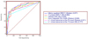 | Figure 2Receiver operating characteristic curves of the best parameters and criteria for discriminating between normal eyes and eyes with early glaucoma using Stratus OCT and 3D OCT. RNFLT = retinal nerve fiber layer thickness; CDR = cup-to-disc ratio.
|
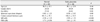
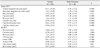
Table 3
AUCs of optic nerve head parameters for discriminating between normal eyes and eyes with early glaucoma using Stratus OCT and 3D OCT
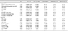
![]()
Table 4
AUCs of retinal nerve fiber layer thickness parameters for discriminating between normal eyes and eyes with early glaucoma using Stratus OCT and 3D OCT
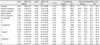
![]()
Table 5
AUCs of criteria based on the normative database for discriminating between normal eyes and eyes with early glaucoma using Stratus OCT and 3D OCT
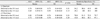
![]()
References
1. Quigley HA, Addicks EM, Green WR. Optic nerve damage in human glaucoma III Quantitative correlation of nerve fiber loss and visual field defect in glaucoma, ischemic neuropathy, papilledema, and toxic neuropathy. Arch Ophthalmol. 1982. 100:135–146.
2. Schuman JS. Spectral domain optical coherence tomography for glaucoma (an AOS thesis). Trans Am Ophthalmol Soc. 2008. 106:426–458.
3. Hood DC, Raza AS, Kay KY, et al. A comparison of retinal nerve fiber layer (RNFL) thickness obtained with frequency and time domain optical coherence tomography (OCT). Opt Express. 2009. 17:3997–4003.
4. Inoue R, Hangai M, Kotera Y, et al. Three-dimensional high-speed optical coherence tomography imaging of lamina cribrosa in glaucoma. Ophthalmology. 2009. 116:214–222.
5. Kotera Y, Hangai M, Hirose F, et al. Three-dimensional imaging of macular inner structures in glaucoma by using spectral-domain optical coherence tomography. Invest Ophthalmol Vis Sci. 2011. 52:1412–1421.
6. Han KE, Jun RM, Choi KR. Comparison of RNFL thickness measured by two different kind of OCT in NTG patients. J Korean Ophthalmol Soc. 2009. 50:1853–1859.
7. Menke MN, Knecht P, Sturm V, et al. Reproducibility of nerve fiber layer thickness measurements using 3D fourier-domain OCT. Invest Ophthalmol Vis Sci. 2008. 49:5386–5391.
8. Hanley JA, McNeil BJ. A method of comparing the areas under receiver operating characteristic curves derived from the same cases. Radiology. 1983. 148:839–843.
9. Cho JW, Sung KR, Hong JT, et al. Detection of glaucoma by spectral domain-scanning laser ophthalmoscopy/optical coherence tomography (SD-SLO/OCT) and time domain optical coherence tomography. J Glaucoma. 2011. 20:15–20.
10. Leung CK, Cheung CY, Weinreb RN, et al. Retinal nerve fiber layer imaging with spectral-domain optical coherence tomography: a variability and diagnostic performance study. Ophthalmology. 2009. 116:1257–1263.
11. Moreno-Montañés J, Olmo N, Alvarez A, et al. Cirrus high-definition optical coherence tomography compared with Stratus optical coherence tomography in glaucoma diagnosis. Invest Ophthalmol Vis Sci. 2010. 51:335–343.
12. Mwanza JC, Oakley JD, Budenz DL, Anderson DR. Cirrus Optical Coherence Tomography Normative Database Study Group. Ability of cirrus HD-OCT optic nerve head parameters to discriminate normal from glaucomatous eyes. Ophthalmology. 2011. 118:241–248.
13. Jeoung JW, Park KH. Comparison of Cirrus OCT and Stratus OCT on the ability to detect localized retinal nerve fiber layer defects in preperimetric glaucoma. Invest Ophthalmol Vis Sci. 2010. 51:938–945.
14. Chang RT, Knight OJ, Feuer WJ, Budenz DL. Sensitivity and specificity of time-domain versus spectral-domain optical coherence tomography in diagnosing early to moderate glaucoma. Ophthalmology. 2009. 116:2294–2299.
15. Sehi M, Grewal DS, Sheets CW, Greenfield DS. Diagnostic ability of Fourier-domain vs time-domain optical coherence tomography for glaucoma detection. Am J Ophthalmol. 2009. 148:597–605.
16. Park SB, Sung KR, Kang SY, et al. Comparison of glaucoma diagnostic Capabilities of Cirrus HD and Stratus optical coherence tomography. Arch Ophthalmol. 2009. 127:1603–1609.
17. Sung KR, Kim DY, Park SB, Kook MS. Comparison of retinal nerve fiber layer thickness measured by Cirrus HD and Stratus optical coherence tomography. Ophthalmology. 2009. 116:1264–1270.
18. Hong S, Seong GJ, Kim SS, et al. Comparison of peripapillary retinal nerve fiber layer thickness measured by spectral vs. time domain optical coherence tomography. Curr Eye Res. 2011. 36:125–134.
19. Leite MT, Rao HL, Zangwill LM, et al. Comparison of the diagnostic accuracies of the Spectralis, Cirrus, and RTVue optical coherence tomography devices in glaucoma. Ophthalmology. 2011. 118:1334–1339.
20. Kim NR, Lee ES, Seong GJ, et al. Spectral-domain optical coherence tomography for detection of localized retinal nerve fiber layer defects in patients with open-angle glaucoma. Arch Ophthalmol. 2010. 128:1121–1128.
21. Budenz DL, Michael A, Chang RT, et al. Sensitivity and specificity of the Stratus OCT for perimetric glaucoma. Ophthalmology. 2005. 112:3–9.
22. Deleón-Ortega JE, Arthur SN, McGwin G Jr, et al. Discrimination between glaucomatous and nonglaucomatous eyes using quantitative imaging devices and subjective optic nerve head assessment. Invest Ophthalmol Vis Sci. 2006. 47:3374–3380.
23. Manassakorn A, Nouri-Mahdavi K, Caprioli J. Comparison of retinal nerve fiber layer thickness and optic disk algorithms with optical coherence tomography to detect glaucoma. Am J Ophthalmol. 2006. 141:105–115.
24. Medeiros FA, Zangwill LM, Bowd C, et al. Evaluation of retinal nerve fiber layer, optic nerve head, and macular thickness measurements for glaucoma detection using optical coherence tomography. Am J Ophthalmol. 2005. 139:44–55.
25. Yüksel N, Altintas O, Ozkan B, et al. Discriminating ability of optical coherence tomography data in staging glaucomatous damage. Can J Ophthalmol. 2009. 44:297–307.
26. Song YM, Uhm KB. Discrimination between normal and early stage of glaucomatous eyes using the Stratus optical coherence tomography. J Korean Ophthalmol Soc. 2007. 48:1675–1685.
27. Brusini P, Salvetat ML, Zeppieri M, et al. Comparison between GDx VCC scanning laser polarimetry and Stratus OCT optical coherence tomography in the diagnosis of chronic glaucoma. Acta Ophthalmol Scand. 2006. 84:650–655.
28. Nouri-Mahdavi K, Hoffman D, Tannenbaum DP, et al. Identifying early glaucoma with optical coherence tomography. Am J Ophthalmol. 2004. 137:228–235.
29. Lee S, Sung KR, Cho JW, et al. Spectral-domain optical coherence tomography and scanning laser polarimetry in glaucoma diagnosis. Jpn J Ophthalmol. 2010. 54:544–549.
30. Rao HL, Zangwill LM, Weinreb RN, et al. Comparison of different spectral domain optical coherence tomography scanning areas for glaucoma diagnosis. Ophthalmology. 2010. 117:1692–1699.
31. Huang JY, Pekmezci M, Mesiwala N, et al. Diagnostic power of optic disc morphology, peripapillary retinal nerve fiber layer thickness, and macular inner retinal layer thickness in glaucoma diagnosis with fourier-domain optical coherence tomography. J Glaucoma. 2011. 20:87–94.
32. Li S, Wang X, Li S, et al. Evaluation of optic nerve head and retinal nerve fiber layer in early and advance glaucoma using frequency-domain optical coherence tomography. Graefes Arch Clin Exp Ophthalmol. 2010. 248:429–434.
33. Aptel F, Sayous R, Fortoul V, et al. Structure-function relationships using spectral-domain optical coherence tomography: comparison with scanning laser polarimetry. Am J Ophthalmol. 2010. 150:825–833.
34. Jonas JB, Fernández MC, Stürmer J. Pattern of glaucomatous neuroretinal rim loss. Ophthalmology. 1993. 100:63–68.
35. Wollstein G, Ishikawa H, Wang J, et al. Comparison of three optical coherence tomography scanning areas for detection of glaucomatous damage. Am J Ophthalmol. 2005. 139:39–43.
36. Kang SM, Lee SB, Uhm KB. Diagnostic ability of Stratus OCT using Korean normative database for early detection of normal-tension glaucoma. J Korean Ophthalmol Soc. 2008. 49:798–810.
37. Sung KR, Kim JS, Wollstein G, et al. Imaging of the retinal nerve fibre layer with spectral domain optical coherence tomography for glaucoma diagnosis. Br J Ophthalmol. 2011. 95:909–914.
 XML Download