

 PDF
PDF ePub
ePub Citation
 Print
Print


Abstract
Methods
Prospectively, 35 patients (70 eyes) were enrolled in the present study. Three sets of corneal curvature values were obtained using the autorefractor (RK-F1®), manual keratometer (OM-2®), partial coherence interferometry keratometer (IOL Master®), wavefront analyzer (KR-1W®), and videokeratography (Orbscan II®). Repeatability of each device was evaluated by coefficient of variation, standard deviation, and intraclass correlation coefficient. RM-ANOVA on ranks was used to compare the differences in corneal curvatures among the devices. The Bland-Altman plot was performed to assess measurement agreement among the devices.
Results
The coefficient of variation values from each device ranged from 2.92% (IOL master®) to 3.06% (Orbscan II®), and the values of intraclass correlation coefficient ranged from 0.965 (KR-1W®) to 0.997 (IOL master®). Compared with the manual keratometer, there was a maximum corneal curvature difference of 1.23 D in KR-1W®, while the other devices had differences less than 0.82 D.
Figures and Tables
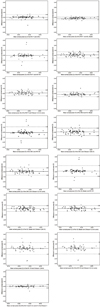 | Figure 1Bland-Altman plots showing differences in corneal curvature measurements between devices. The upper and lower dashed lines indicate 95% limits of agreement and the solid line indicates the mean.
|
Table 1
Results for repeatability of seven corneal curvature measurements by coefficient of variation, standard deviation, and intraclass correlation coefficient

![]()
Table 2
Results for the comparison of the 6 corneal curvature measurements among devices
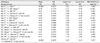
The mean difference, SD, upper LoA, lower LoA, RM-ANOVA on Ranks are displayed for each pair of devices.
D = diopters; SD = standard deviation; LoA = 95% limits of agreement; RM-ANOVA on Ranks = repeated measures analysis of variance on ranks; 15 comparisons were made for each device pair (3 measurements each).
![]()
References
1. Olsen T. Sources of error in intraocular lens power calculation. J Cataract Refract Surg. 1992. 18:125–129.
2. Maeng HS, Ryu EH, Chung TY, Chung ES. Effects of anterior chamber depth and axial length on refractive error after intraocular lens implantation. J Korean Ophthalmol Soc. 2010. 51:195–202.
3. Koranyi G, Lydahl E, Norrby S, Taube M. Anterior chamber depth measurement: a-scan versus optical methods. J Cataract Refract Surg. 2002. 28:243–247.
4. Hosny M, Alio JL, Claramonte P, et al. Relationship between anterior chamber depth, refractive state, corneal diameter, and axial length. J Refract Surg. 2000. 16:336–340.
5. Olsen T. Prediction of the effective postoperative (intraocular lens) anterior chamber depth. J Cataract Refract Surg. 2006. 32:419–424.
6. Norrby S. Sources of error in intraocular lens power calculation. J Cataract Refract Surg. 2008. 34:368–376.
7. Qazi MA, Cua IY, Roberts CJ, Pepose JS. Determining corneal power using Orbscan II videokeratography for intraocular lens calculation after excimer laser surgery for myopia. J Cataract Refract Surg. 2007. 33:21–30.
8. Elbaz U, Barkana Y, Gerber Y, et al. Comparison of different techniques of anterior chamber depth and keratometric measurements. Am J Ophthalmol. 2007. 143:48–53.
9. Elliott M, Simpson T, Richter D, Fonn D. Repeatability and accuracy of automated refraction: a comparison of the Nikon NRK-8000, the Nidek AR-1000, and subjective refraction. Optom Vis Sci. 1997. 74:434–438.
10. Holzer MP, Mamusa M, Auffarth GU. Accuracy of a new partial coherence interferometry analyser for biometric measurements. Br J Ophthalmol. 2009. 93:807–810.
11. Choi JH, Roh GH. The reproducibility and accuracy of biometry parameter measurement from IOL Master®. J Korean Ophthalmol Soc. 2004. 45:1665–1673.
12. Findl O, Drexler W, Menapace R, et al. Improved prediction of intraocular lens power using partial coherence interferometry. J Cataract Refract Surg. 2001. 27:861–867.
13. Findl O, Drexler W, Menapace R, et al. High precision biometry of pseudophakic eyes using partial coherence interferometry. J Cataract Refract Surg. 1998. 24:1087–1093.
14. Speicher L. Intra-ocular lens calculation status after corneal refractive surgery. Curr Opin Ophthalmol. 2001. 12:17–29.
15. Holladay JT, Prager TC, Ruiz RS, et al. Improving the predictability of intraocular lens power calculations. Arch Ophthalmol. 1986. 104:539–541.
16. Mamalis N. Complications of foldable intraocular lenses requiring explanation or secondary intervention--1998 survey. J Cataract Refract Surg. 2000. 26:766–772.
17. Shin YJ, Kim NH, Kim DH. Comparison of pentacam with Orbscan. J Korean Ophthalmol Soc. 2007. 48:637–641.
18. Park SJ, Wee WR, Lee JH, Kim MK. Comparison of wavescan aberrometer refraction to subjective manifest refraction and autorefractor. J Korean Ophthalmol Soc. 2009. 50:684–690.
19. Jo DH, Oh JY, Kim MK, et al. Corneal power estimation using Orbscan II videokeratography in eyes with previous corneal refractive surgeries. J Korean Ophthalmol Soc. 2009. 50:1730–1734.
20. Solomon KD, Fernandez de Castro LE, Sandoval HP, Vroman DT. Comparison of wavefront sensing devices. Ophthalmol Clin North Am. 2004. 17:119–127.
21. Cairns G, McGhee CN. Orbscan computerized topography: attributes, applications, and limitations. J Cataract Refract Surg. 2005. 31:205–220.
22. Menassa N, Kaufmann C, Goggin M, et al. Comparison and reproducibility of corneal thickness and curvature readings obtained by the Galilei and the Orbscan II analysis systems. J Cataract Refract Surg. 2008. 34:1742–1747.
23. Kawamorita T, Uozato H, Kamiya K, et al. Repeatability, reproducibility, and agreement characteristics of rotating Scheimpflug photography and scanning-slit corneal topography for corneal power measurement. J Cataract Refract Surg. 2009. 35:127–133.
24. Shirayama M, Wang L, Weikert MP, Koch DD. Comparison of corneal power obtained from 4 different devices. Am J Ophthalmol. 2009. 148:528–535.
25. Huynh SC, Mai TQ, Kifley A, et al. An evaluation of keratometry in 6-year-old children. Cornea. 2006. 25:383–387.
26. Kiely PM, Smith G, Carney LG. Meridional variations of corneal shape. Am J Optom Physiol Opt. 1984. 61:619–626.
27. Hayashi K, Hayashi H, Hayashi F. Topographic analysis of the changes in corneal shape due to aging. Cornea. 1995. 14:527–532.
28. Kim CS, Kim SY, Park YH, Lee YC. Change in ocular dimensions with age in patients with emmetropia. J Korean Ophthalmol Soc. 2008. 49:425–432.
 XML Download