

 PDF
PDF ePub
ePub Citation
 Print
Print


Abstract
Purpose
To evaluate the response and cellular damage of cultured human pterygial cells according to the concentration and exposure time of topical cyclosporin.
Methods
Human pterygial cells were exposed to a cyclosporin A concentrations of 0.1 µg/ml (0.0001%), 1 µg/ml (0.0001%), 10 µg/ml (0.001%), 100 µg/ml (0.01%), or 500 µg/ml (0.05%) for 5 or 10 minutes. An MTT-based colorimetric assay was performed to assess the metabolic activity of cellular proliferation, and a lactate dehydrogenase (LDH) leakage assay was used to determine cellular damage. The extra-cellular matrix of PIP, laminin and MMP were evaluated, and the measurement of pro-inflammatory cytokine, TNF-α and IL-1b. IL-6, IL-8 was performed using ELISA kits.
Results
The pterygial cellular inhibitory effect of cyclosporin was similar to that of the control according to the concentration and exposure time (p > 0.05). Compared with the control, the level of LDH did not show a statistically significant difference between concentration and exposure time (p > 0.05). There was no significant difference of inhibitory effects by PIP, laminin, or MMP between the experimental and control groups (p > 0.05). The production of TNF-α and IL from the experimental pterygial cells due to the effect of cyclosporin was not significantly different from that of the control at a longer exposure time or stronger concentration (p > 0.05).
Conclusions
The response of pterygial cells to topical cyclosporin A at concentrations less than 0.05% for less than 10 minutes of exposure time showed no prevention of pterygial recurrence. With regard to cellular damage, little effects on inhibition of PIP, laminin, MMP, IL, and TNF-α were observed compared with that of the control.
Figures and Tables
Figure 1
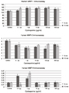
Metabolic activities of cultured human pterygial cells measured with MTT assays by a scanning spectrometer (ELISA reader). There is no significant difference of cellular viabilities between the experimental and control groups with increasing concentration and longer exposure time (p > 0.05).
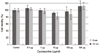
Figure 2
LDH titers of cultured human pterygial cells exposed in cyclosporine. There is no significant difference of LDH activities in cultured pterygial cells between the experimental and control groups according to concentration and exposure time (p > 0.05).
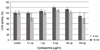
Figure 3
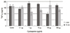
PIP level of cultured human pterygial cells exposed in cyclosporine. There is no significant difference of PIP levels in cultured pterygial cells between the experimental and control groups according to concentration and exposure time (p > 0.05).
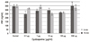
Figure 4
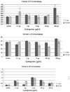
Laminin level of cultured human pterygial cells exposed to cyclosporin. There is no significant difference of laminin levels in cultured pterygial cells between the experimental and control groups according to concentration and exposure time (p > 0.05).
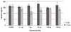
Figure 5
MMP level of cultured human pterygial cells exposed to cyclosporine. There is no significant difference of MMP-1 (upper), MMP-2 (middle), MMP-9 (lower) levels in cultured pterygial cells between the experimental and control groups according to concentration and exposure time (p > 0.05).
References
1. Coroneo MT, Di Girolamo N, Wakefield D. The pathogenesis of pterygia. Curr Opin Ophthalmol. 1999. 10:282–288.
2. Hill JC, Maske R. Pathogenesis of pterygium. Eye (Lond). 1989. 3:218–226.
3. Cameron ME. Histology of pterygium: an electron microscopic study. Br J Ophthalmol. 1983. 67:604–608.
4. Chan CM, Liu YP, Tan DT. Ocular surface changes in pterygium. Cornea. 2002. 21:38–42.
5. Wang IJ, Lai WT, Liou SW. Impression cytology of pterygium. J Ocul Pharmacol Ther. 2000. 16:519–528.
6. Di Girolamo N, Chui J, Coroneo MT, Wakefield D. Pathogenesis of pterygia: role of cytokines, growth factors, and matrix metalloproteinases. Prog Retin Eye Res. 2004. 23:195–228.
7. Baudouin C, Fredj-Reygrobellet D, Gastaud P, Lapalus P. HLA DR and DQ distribution in normal human ocular structures. Curr Eye Res. 1988. 7:903–911.
8. Byun YS, Jeon EJ, Chung SK. Clinical effect of cyclosporine 0.05% eye drops in dry eye syndrome patients. J Korean Ophthalmol Soc. 2008. 49:1583–1588.
9. Di Girolamo N, Kumar RK, Coroneo MT, Wakefield D. UVB-mediated induction of interleukin-6 and -8 in pterygia and cultured human pterygium epithelial cells. Invest Ophthalmol Vis Sci. 2002. 43:3430–3437.
10. Strong B, Farley W, Stern ME, Pflugfelder SC. Topical cyclosporine inhibits conjunctival epithelial apoptosis in experimental murine keratoconjunctivitis sicca. Cornea. 2005. 24:80–85.
11. Barry A. Schechter. Cyclosporine may be new treatment option for pterygia; Offers alternative to topical corticosteroids, may reduce or delay need for surgery. Ophthalmology Times. 2007. 6:33–34.
12. Turan-Vural E, Torun-Acar B, Kivanc SA, Acar S. The effect of topical 0.05% cyclosporine on recurrence following pterygium surgery. Clin Ophthalmol. 2011. 5:881–885.
13. Yalcin Tok O, Burcu Nurozler A, Ergun G, et al. Topical cyclosporine A in the prevention of pterygium recurrence. Ophthalmologica. 2008. 222:391–396.
14. Ibáñez M, Eugarrios MF, Calderón DI. Topical cyclosporin A and mitomycin C injection as adjunctive therapy for prevention of primary pterygium recurrence. Ophthalmic Surg Lasers Imaging. 2009. 40:239–244.
15. Song YS, Lee JK, Kim JC. Circulating endothelial progenitor cell and vasculogenic factors in pterygium pathogenesis. J Korean Ophthalmol Soc. 2006. 47:1472–1480.
16. Mauger TF, Craig EL, editors. Havener's Ocular Pharmacology. 1994. 6th ed. St. Louis: Mosby;chap. 2.
17. Shin JH, Kim SJ, Lee JE, Lee JS. Effect of mitomycin C, dexamethasone, and cyclosporin A 0.05% on the proliferation of human corneal ketatocyte. J Korean Ophthalmol Soc. 2011. 52:1215–1221.
18. Lee JS, Oum BS, Lee SH. Mitomycin C influence on inhibition of cellular proliferation and subsequent synthesis of type I collagen and laminin in primary and recurrent pterygia. Ophthalmic Res. 2001. 33:140–146.
19. Berman B, Duncan MR. Pentoxifylline inhibits normal human dermal fibroblast in vitro proliferation, collagen, glycosaminoglycan, and fibronectin production, and increases collagenase activity. J Invest Dermatol. 1989. 92:605–610.
20. Leonardi A, DeFranchis G, Fregona IA, et al. Effects of cyclosporin A on human conjunctival fibroblasts. Arch Ophthalmol. 2001. 119:1512–1517.
21. Bankers-Fulbright JL, Kalli KR, McKean DJ. Interleukin-1 signal transduction. Life Sci. 1996. 59:61–83.
22. Power WJ, Mullaney P, Farrell M, Collum LM. Effect of topical cyclosporin A on conjunctival T cells in patients with secondary Sjögren's syndrome. Cornea. 1993. 12:507–511.
23. Kunert KS, Tisdale AS, Stern ME, et al. Analysis of topical cyclosporine treatment of patients with dry eye syndrome: effect on conjunctival lymphocytes. Arch Ophthalmol. 2000. 118:1489–1496.
24. Oh C, Apel AJ, Saville BA, et al. Local efficacy of cyclosporine in corneal transplant therapy. Curr Eye Res. 1994. 13:337–343.
25. Occleston NL, Alexander RA, Mazure A, et al. Effects of single exposures to antiproliferative agents on ocular fibroblast-mediated collagen contraction. Invest Ophthalmol Vis Sci. 1994. 35:3681–3690.
 XML Download