

 PDF
PDF ePub
ePub Citation
 Print
Print


Abstract
Purpose
To compare postoperative clinical outcomes between 2 types of 3-piece aspheric intraocular lenses.
Methods
Uncorrected visual acuity, best corrected visual acuity, total ocular and internal ocular aberration including higher-order aberrations and spherical aberration, and modulation transfer functions were compared 6 months after cataract surgery between eyes implanted with TECNIS ZA9003 (group 1) and HOYA PC-60AD (group 2) in 30 and 28 eyes, respectively, In addition, the differences between postoperative spherical equivalent and preoperative target refractive errors were analyzed.
Results
Clinical outcomes showed no significant differences between both groups including visual acuities, high order aberrations, and modulation transfer function. In both groups, postoperative refractive errors were more of a myopic state than preoperative estimated target refraction. The myopic refractive error between both groups showed no significant difference (-0.26 vs. -0.42 diopter, p = 0.75).
Figures and Tables


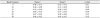
Table 4
Total ocular aberrations (µm) between the groups measured by iTrace® 6 months after operation


References
1. Werner L, Olson RJ, Mamalis N. New technology IOL optics. Ophthalmol Clin North Am. 2006. 19:469–483.
2. Holladay JT, Piers PA, Koranyi G, et al. A new intraocular lens design to reduce spherical aberration of pseudophakic eyes. J Refract Surg. 2002. 18:683–691.
3. McLellan JS, Marcos S, Burns SA. Age-related changes in monochromatic wave aberrations of the human eye. Invest Ophthalmol Vis Sci. 2001. 42:1390–1395.
4. Oshika T, Klyce SD, Applegate RA, Howland HC. Changes in corneal wavefront aberrations with aging. Invest Ophthalmol Vis Sci. 1999. 40:1351–1355.
5. Artal P, Guirao A, Berrio E, Williams DR. Compensation of corneal aberrations by the internal optics in the human eye. J Vis. 2001. 1:1–8.
6. Applegate RA, Howland HC, Sharp RP, et al. Corneal aberrations and visual performance after radial keratotomy. J Refract Surg. 1998. 14:397–407.
7. Rawer R, Stork W, Spraul CW, Lingenfelder C. Imaging quality of intraocular lenses. J Cataract Refract Surg. 2005. 31:1618–1631.
8. Guirao A, Redondo M, Geraghty E, et al. Corneal optical aberrations and retinal image quality in patients in whom monofocal intraocular lenses were implanted. Arch Ophthalmol. 2002. 120:1143–1151.
9. Altmann GE, Nichamin LD, Lane SS, Pepose JS. Optical performance of 3 intraocular lens designs in the presence of decentration. J Cataract Refract Surg. 2005. 31:574–585.
10. Caporossi A, Martone G, Casprini F, Rapisarda L. Prospective randomized study of clinical performance of 3 aspheric and 2 spherical intraocular lenses in 250 eyes. J Refract Surg. 2007. 23:639–648.
11. Mester U, Dillinger P, Anterist N. Impact of a modified optic design on visual function: clinical comparative study. J Cataract Refract Surg. 2003. 29:652–660.
12. Ahn HS, Kim SW, Kim EK, Kim TI. Wavefront and visual function analysis after aspherical and spherical intraocular lenses implantation. J Korean Ophthalmol Soc. 2008. 49:1248–1255.
13. Oshika T, Nagata T, Ishii Y. Adhesion of lens capsule to intraocular lenses of polymethylmethacrylate, silicone, and acrylic foldable materials: an experimental study. Br J Ophthalmol. 1998. 82:549–553.
14. Versura P, Torreggiani A, Cellini M, Caramazza R. Adhesion mechanisms of human lens epithelial cells on 4 intraocular lens materials. J Cataract Refract Surg. 1999. 25:527–533.
15. Nejima R, Miyata K, Honbou M, et al. A prospective, randomised comparison of single and three piece acrylic foldable intraocular lenses. Br J Ophthalmol. 2004. 88:746–749.
16. Hayashi K, Hayashi H. Comparison of the stability of 1-piece and 3-piece acrylic intraocular lenses in the lens capsule. J Cataract Refract Surg. 2005. 31:337–342.
17. Son SW, Seo JW, Shin SJ, Chung SK. Comparison of the stability between three-piece and single-piece aspheric intraocular lenses. J Korean Ophthalmol Soc. 2010. 51:1584–1589.
18. Ohtani S, Miyata K, Samejima T, et al. Intraindividual comparison of aspherical and spherical intraocular lenses of same material and platform. Ophthalmology. 2009. 116:896–901.
19. Yamaguchi T, Negishi K, Ono T, et al. Feasibility of spherical aberration correction with aspheric intraocular lenses in cataract surgery based on individual pupil diameter. J Cataract Refract Surg. 2009. 35:1725–1733.
20. Kim SW, Ahn H, Kim EK, Kim TI. Comparison of higher order aberrations in eyes with aspherical or spherical intraocular lenses. Eye (Lond). 2008. 22:1493–1498.
21. Takeo S, Watanabe Y, Suzuki M, Kadonosono K. Wavefront analysis of acrylic spherical and aspherical intraocular lenses. Jpn J Ophthalmol. 2008. 52:250–254.
22. Wang L, Dai E, Koch DD, Nathoo A. Optical aberrations of the human anterior cornea. J Cataract Refract Surg. 2003. 29:1514–1521.
23. Beiko GH, Haigis W, Steinmueller A. Distribution of corneal spherical aberration in a comprehensive ophthalmology practice and whether keratometry can predict aberration values. J Cataract Refract Surg. 2007. 33:848–858.
24. Levy Y, Segal O, Avni I, Zadok D. Ocular higher-order aberrations in eyes with supernormal vision. Am J Ophthalmol. 2005. 139:225–228.
25. Norrby NE, Grossman LW, Geraghty EP, et al. Determining the imaging quality of intraocular lenses. J Cataract Refract Surg. 1998. 24:703–714.
 XML Download