

 PDF
PDF ePub
ePub Citation
 Print
Print


Abstract
Purpose
To compare the changes in central subfield macular thickness (CSMT) using optical coherence tomography (OCT) after cataract surgery and to evaluate the risk factors of macular edema.
Methods
The retrospective study consisted of 55 eyes of 50 patients who underwent phacoemulsification. All of these eyes were assessed by OCT and best-corrected visual acuity, and fundus examination before and one month after surgery. CSMT was measured based on a central thickness of 1 mm, and macular edema was defined as an increase of CSMT in 30% or more after surgery than before.
Results
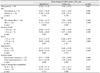
CSMT significantly increased by 22.2 ± 47.10 µm at 1 month after cataract surgery in the operated eye compared with the fellow eye (p = 0.01). The diabetic retinopathy group and hypertensive group showed more significant increases than the non-diabetic and non-hypertensive group. Macular edema developed in seven of 55 eyes (12.7%), and they consisted of five moderate or more severe diabetic retinopathies and two epiretinal membrane. Four weeks after surgery, the macular edema group showed more decrease in visual acuity; however, the CSMT was not correlated with the duration of diabetes mellitus, types of surgical incision, HbA1c or other factors.
Figures and Tables
Figure 1
(A) Preoperative fundus photograph shows severe nonproliferative diabetic retinopathy and hard exudates near macula. OCT image shows normal foveal contour. (B) Postoperative fundus photograph and OCT image shows significant macular edema.
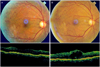
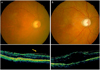
Figure 2
(A) Preoperative fundus photograph shows epiretinal membreane. (B) Postoperative OCT image shows epiretinal membrane and significant macular edema.


References
1. Rossetti L, Autelitano A. Cystoid macular edema following cataract surgery. Curr Opin Ophthalmol. 2000. 11:65–72.
2. Rossetti L, Chaudhuri J, Dickersin K. Medical prophylaxis and treatment of cystoid macular edema after cataract surgery: The results of a meta-analysis. Ophthalmology. 1998. 105:397–405.
3. Norregaard JC, Bernth-Petersen P, Bellan L, et al. Intraoperative clinical practice and risk of early complications after cataract extraction in the United States, Canada, Denmark, and Spain. Ophthalmology. 1999. 106:42–48.
4. Powe NR, Schein OD, Gieser SC, et al. Synthesis of the literature on visual acuity and complications following cataract extraction with intraocular lens implantation. Cataract Patient Outcome Research Team. Arch Ophthalmol. 1994. 112:239–252.
5. Riley AF, Malik TY, Grupcheva CN, et al. The Auckland cataract study: co-morbidity, surgical techniques, and clinical outcomes in a public hospital service. Br J Ophthalmol. 2002. 86:185–190.
6. Kim SJ, Equi R, Bressler NM. Analysis of macular edema after cataract surgery in patients with diabetes using optical coherence tomography. Ophthalmology. 2007. 114:881–889.
7. Kim JY, Song MH, Chung SK. Analysis of postoperative macular edema in cataract patients with diabetes using optical coherence tomography. J Korean Ophthalmol Soc. 2010. 51:340–346.
8. Wang SJ, Choi SH. The changes in macular thickness after phacoemulsification in patients with non-diabetes and nonproliferative diabetic retinopathy. J Korean Ophthalmol Soc. 2008. 49:57–64.
9. Perente I, Utine CA, Ozturker C, et al. Evaluation of macular changes after uncomplicated phacoemulsification surgery by optical coherence tomography. Curr Eye Res. 2007. 32:241–247.
10. Kim SJ, Belair ML, Bressler NM, et al. A method of reporting macular edema after cataract surgery using optical coherence tomography. Retina. 2008. 28:870–876.
11. Biro Z, Balla Z, Kovacs B. Change of foveal and perifoveal thickness measured by OCT after phacoemulsification and IOL implantation. Eye (Lond). 2008. 22:8–12.
12. Dowler JG, Sehmi KS, Hykin PG, Hamilton AM. The natural history of macular edema after cataract surgery in diabetes. Ophthalmology. 1999. 106:663–668.
13. Nussenblatt RB, Kaufman SC, Palestine AG, et al. Macular thickening and visual acuity. Measurement in patients with cystoid macular edema. Ophthalmology. 1987. 94:1134–1139.
14. Hee MR, Puliafito CA, Duker JS, et al. Topography of diabetic macular edema with optical coherence tomography. Ophthalmology. 1998. 105:360–370.
15. Kim SJ, Bressler NM. Optical coherence tomography and cataract surgery. Curr Opin Ophthalmol. 2009. 20:46–51.
16. Estafanous MF, Lowder CY, Meisler DM, Chauhan R. Phacoemulsification cataract extraction and posterior chamber lens implantation in patients with uveitis. Am J Ophthalmol. 2001. 131:620–625.
17. Quinn CJ. Cystoid macular edema. Optom Clin. 1996. 5:111–130.
18. Tso MO. Pathology of cystoid macular edema. Ophthalmology. 1982. 89:902–915.
19. Krishna R, Meisler DM, Lowder CY, et al. Long-term follow-up of extracapsular cataract extraction and posterior chamber intraocular lens implantation in patients with uveitis. Ophthalmology. 1998. 105:1765–1769.
20. Torrón-Fernández-Blanco C, Ruiz-Moreno O, Ferrer-Novella E, et al. Pseudophakic cystoid macular edema. Assessment with optical coherence tomography. Arch Soc Esp Oftalmol. 2006. 81:147–153.
21. Nicholas S, Riley A, Patel H, et al. Correlations between optical coherence tomography measurement of macular thickness and visual acuity after cataract extraction. Clin Experiment Ophthalmol. 2006. 34:124–129.
22. Ching HY, Wong AC, Wong CC, et al. Cystoid macular oedema and changes in retinal thickness after phacoemulsification with optical coherence tomography. Eye (Lond). 2006. 20:297–303.
23. Biró Z, Balla Z. OCT measurements on the foveal and perifoveal retinal thickness on diabetic patients after phacoemulsification and IOL implantation. Eye (Lond). 2010. 24:639–647.
24. Eriksson U, Alm A, Bjärnhall G, et al. Macular edema and visual outcome following cataract surgery in patients with diabetic retinopathy and controls. Graefes Arch Clin Exp Ophthalmol. 2011. 249:349–359.
25. Hayashi K, Igarashi C, Hirata A, Hayashi H. Changes in diabetic macular oedema after phacoemulsification surgery. Eye (Lond). 2009. 23:389–396.
26. Mackool RJ, Muldoon T, Fortier A, Nelson D. Epinephrine-induced cystoid macular edema in aphakic eyes. Arch Ophthalmol. 1977. 95:791–793.
27. Michels RG, Maumenee AE. Cystoid macular edema associated with topically applied epinephrine in aphakic eyes. Am J Ophthalmol. 1975. 80:379–388.
28. Classé JG. Epinephrine maculopathy. J Am Optom Assoc. 1980. 51:1091–1093.
29. Thomas JV, Gragoudas ES, Blair NP, Lapus JV. Correlation of epinephrine use and macular edema in aphakic glaucomatous eyes. Arch Ophthalmol. 1978. 96:625–628.
30. Bozkurt E, Yazici AT, Pekel G, et al. Effect of intracameral epinephrine use on macular thickness after uneventful phacoemulsification. J Cataract Refract Surg. 2010. 36:1380–1384.
31. Jurecka T, Bátková Z, Ventruba J, Synek S. Macular edema after cataract surgery in diabetic patients without retinopathy. Cesk Slov Oftalmol. 2007. 63:274–284.
32. Ghosh S, Roy I, Biswas PN, et al. Prospective randomized comparative study of macular thickness following phacoemulsification and manual small incision cataract surgery. Acta Ophthalmol. 2010. 88:e102–e106.
33. Jain R, Stevens JD, Bunce CV, et al. Ischaemic heart disease may predispose to pseudophakic cystoid macular oedema. Eye (Lond). 2001. 15:34–38.
34. Henderson BA, Kim JY, Ament CS, et al. Clinical pseudophakic cystoid macular edema. Risk factors for development and duration after treatment. J Cataract Refract Surg. 2007. 33:1550–1558.
35. Martinez MR, Ophir A. Pseudophakic cystoid macular edema associated with extrafoveal vitreoretinal traction. Open Ophthalmol J. 2011. 5:35–41.
36. Pollack A, Leiba H, Bukelman A, et al. The course of diabetic retinopathy following cataract surgery in eyes previously treated by laser photocoagulation. Br J Ophthalmol. 1992. 76:228–231.
 XML Download