

 PDF
PDF ePub
ePub Citation
 Print
Print


Abstract
Purpose
To evaluate long-term endothelial cell changes in phakic eyes that underwent implantation of an angle-supported anterior chamber lens to correct myopia.
Methods
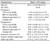
A retrospective analysis was performed in 110 eyes of 55 patients who underwent implantation of angle-supported anterior chamber lenses with a follow-up period longer than 5 years. Comparisons were made between preoperative and postoperative endothelial cell density, coefficient of variation, and percentage of hexagonal cells.
Results
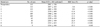
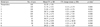
Mean preoperative corneal endothelial cell density was 2951 ± 336 cells/mm2 and the percentage of cell loss was 3.8% at year 1, 12.6% at year 3, 13.4% at year 5, 22.5% at year 7, and 22.2% at year 9. Explantation was required in 13 eyes (11.8%) due to the decrease of endothelial cell count to 936 ± 458 cells/mm2 over 9 years of follow-up.
Figures and Tables
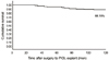
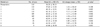
Figure 1
Phakic intraocular lens (PIOL) explantation. Kaplan-Meier survival table. After 112 months of follow-up, 88.18% of cases will retain the PIOL in place.
References
1. Seiler T, Holschbach A, Derse M, et al. Complications of myopic photorefractive keratectomy with the excimer laser. Ophthalmology. 1994. 101:153–160.
2. Binder PS. Ectasia after laser in situ keratomileusis. J Cataract Refract Surg. 2003. 29:2419–2429.
3. Pallikaris IG, Kymionis GD, Astyrakakis NI. Corneal ectasia induced by laser in situ keratomileusis. J Cataract Refract Surg. 2001. 27:1796–1802.
4. Teus MA, de Benito-Llopis L, García-González M. Comparison of visual results between laser-assisted subepithelial keratectomy and epipolis laser in situ keratomileusis to correct myopia and myopic astigmatism. Am J Ophthalmol. 2008. 146:357–362.
5. Javitt JC. Clear-lens extraction for high myopia. Is this an idea whose time has come? Arch Ophthalmol. 1994. 112:321–323.
6. Saragoussi JJ, Cotinat J, Renard G, et al. Damage to the corneal endothelium by minus power anterior chamber intraocular lenses. Refract Corneal Surg. 1991. 7:282–285.
7. Mimouni F, Colin J, Koffi V, Bonnet P. Damage to the corneal endothelium from anterior chamber intraocular lenses in phakic myopic eyes. Refract Corneal Surg. 1991. 7:277–281.
8. Pérez-Santonja JJ, Iradier MT, Sanz-Iglesias L, et al. Endothelial changes in phakic eyes with anterior chamber intraocular lenses to correct high myopia. J Cataract Refract Surg. 1996. 22:1017–1022.
9. Kohnen T, Kook D, Morral M, Güell JL. Phakic intraocular lenses: part 2: results and complications. J Cataract Refract Surg. 2010. 36:2168–2194.
10. Huang D, Schallhorn SC, Sugar A, et al. Phakic intraocular lens implantation for the correction of myopia. Ophthalmology. 2009. 116:2244–2258.
11. Javaloy J, Alió JL, Iradier MT, et al. Outcomes of ZB5M angle-supported anterior chamber phakic intraocular lenses at 12 years. J Refract Surg. 2007. 23:147–158.
12. Doors M, Cals D, Berendschot T, et al. Influence of anterior chamber morphometrics on endothelial cell changes after phakic intraocular lens implantation. J Cataract Refract Surg. 2008. 34:2110–2118.
13. Engelmann K, Bednarz J, Valtink M. Prospects for endothelial transplantation. Exp Eye Res. 2004. 78:573–578.
14. Joyce NC. Cell cycle status in human corneal endothelium. Exp Eye Res. 2005. 81:629–638.
15. Güell JL, Morral M, Gris O, et al. Five-year follow-up of 399 phakic Artisan-Verisyse implantation for myopia, hyperopia, and/or astigmatism. Ophthalmology. 2008. 115:1002–1012.
16. Waring GO 3rd, Bourne WM, Edelhauser HF, Kenyon KR. The corneal endothelium. Normal and pathologic structure and function. Ophthalmology. 1982. 89:531–590.
17. Yee RW, Matsuda M, Schultz RO, Edelhauser HF. Changes in the normal corneal endothelial cellular pattern as a function of age. Curr Eye Res. 1985. 4:671–678.
18. Benedetti S, Casamenti V, Benedetti M. Long-term endothelial changes in phakic eyes after Artisan intraocular lens implantation to correct myopia. J Cataract Refract Surg. 2007. 33:784–790.
19. Knorz MC, Lane SS, Holland SP. Angle-supported phakic intraocular lens for correction of moderate to high myopia: Three-year interim results in international multicenter studies. J Cataract Refract Surg. 2011. 37:469–480.
20. Allemann N, Chamon W, Tanaka HM, et al. Myopic angle-supported intraocular lenses: two-year follow-up. Ophthalmology. 2000. 107:1549–1554.
21. Pérez-Santonja JJ, Alió JL, Jiménez-Alfaro I, Zato MA. Surgical correction of severe myopia with an angle-supported phakic intraocular lens. J Cataract Refract Surg. 2000. 26:1288–1302.
22. Leccisotti A, Fields SV. Clinical results of ZSAL-4 angle-supported phakic intraocular lenses in 190 myopic eyes. J Cataract Refract Surg. 2005. 31:318–323.
23. Alió JL, Pinero D, Bernabeu G, et al. The Kelman Duet phakic intraocular lens: 1-year results. J Refract Surg. 2007. 23:868–879.
24. Stulting RD, John ME, Maloney RK, et al. Three-year results of Artisan/Verisyse phakic intraocular lens implantation. Results of the United States Food and Drug Administration clinical trial. Ophthalmology. 2008. 115:464–472.
25. Budo C, Hessloehl JC, Izak M, et al. Muticenter study of the Artisan phakic intraocular lens. J Cataract Refract Surg. 2000. 26:1163–1171.
26. Landesz M, van Rij G, Luyten G. Iris-claw phakic intraocular lens for high myopia. J Refract Surg. 2001. 17:634–640.
27. Menezo JL, Avino JA, Cisneros A, et al. Iris-claw phakic intraocular lens for high myopia. J Refract Surg. 1997. 13:545–555.
28. Pérez-Santonja JJ, Bueno JL, Zato MA. Surgical correction of high myopia in phakic eyes with Worst-Fechner myopia intraocular lenses. J Refract Surg. 1997. 13:268–281.
29. Silva RA, Jain A, Manche EE. Prospective long-term evaluation of the efficacy, safety, and stability of the phakic intraocular lens for high myopia. Arch Ophthalmol. 2008. 126:775–781.
30. Bourne WM, Nelson LR, Hodge DO. Central corneal endothelial cell changes over a ten-year period. Invest Ophthalmol Vis Sci. 1997. 38:779–782.
31. Jang BH, Lee DW, Cho NC, Ahn M. Clinical results of anterior chamber phakic intraocular lens. J Korean Ophthalmol Soc. 2006. 47:31–36.
32. Alió JL, de la Hoz F, Pérez-Santonja JJ, et al. Phakic anterior chamber lenses for the correction of myopia: a 7-year cumulative analysis of complications in 263 cases. Ophthalmology. 1999. 106:458–466.
33. Baikoff G. The refractive IOL in a phakic eye. Ophthalmic Practice. 1991. 9:58–61.
34. Alió JL, Abdelrahman AM, Javaloy J, et al. Angle-supported anterior chamber phakic intraocular lens explantation causes and outcome. Ophthalmology. 2006. 113:2213–2220.
35. Coullet J, Mahieu L, Malecaze F, et al. Severe endothelial cell loss following uneventful angle-supported phakic intraocular lens implantation for high myopia. J Cataract Refract Surg. 2007. 33:1477–1481.
36. Kim M, Kim JK, Lee HK. Corneal endothelial decompensation after iris-claw phakic intraocular lens implantation. J Cataract Refract Surg. 2008. 34:517–519.
37. Saxena R, Boekhoorn S, Mulder P, et al. Long-term follow-up of endothelial cell change after Artisan phakic intraocular lens implantation. Ophthalmology. 2008. 115:608–613.
 XML Download