

 PDF
PDF ePub
ePub Citation
 Print
Print


Abstract
Purpose
To evaluate the difference between corneal endothelial cell density at the moment of preservation and at keratoplasty in imported donor corneas and to analyze the correlated factors of the difference.
Methods
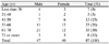
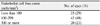
Eighty-seven imported corneas were evaluated. Corneal endothelial cell density at the moment of preservation was obtained from the medical record and was measured just before the keratoplasty. Correlation of the difference in endothelial cell density with the following factors were analyzed; donor sex, donor age, death-to-preservation time, preservation-to-surgery time, death-to-surgery time, endothelial cell density at the moment of preservation, and preservation period of the corneas.
Results
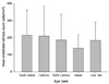
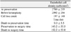
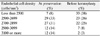
All of the corneas showed a decrease in endothelial cell density. Mean endothelial cell density of imported donor corneas at the moment of preservation and at keratoplasty was 2789 ± 235 cells/mm2 and 2592 ± 254 cells/mm2 (p < 0.001), respectively. Mean endothelial cell loss was 197 ± 148 cells/mm2, which was significantly correlated with preservation-to-surgery time, death-to-surgery time and a preservation period longer than 7 days (p = 0.042, p = 0.045, p = 0.036, respectively).
Figures and Tables
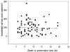 | Figure 3Correlation between death to preservation time and endothelial cell loss count (r = -0.040, p = 0.714). Mean death to preservation time was 9.0 ± 5.5 hours.
|
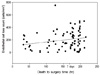 | Figure 4Correlation between death to surgery time and endothelial cell loss count (r = 0.216, p = 0.045). Mean death to surgery time was 152.2 ± 53.0 hours (6.34 days).
|

References
1. Barron BA. Kaufman HE, Barron BA, McDonald MB, editors. Penetrating keratoplasty. The Cornea. 1998. 2nd ed. Boston: Butterworth-Heinemann;chap. 34.
2. Krohn J, Høvding G. The influence of donor age and cause of death on corneal endothelial cell density. Acta Ophthalmol Scand. 2005. 83:746–750.
3. Williams KA, Roder D, Esterman A, et al. Factors predictive of corneal graft survival. Report from the Australian Corneal Graft Registry. Ophthalmology. 1992. 99:403–414.
4. Ha DW, Kim CK, Lee SE, et al. Penet rating keratoplasty results in 275 cases. J Korean Ophthalmol Soc. 2001. 42:20–29.
5. Polack FM. The endothelium of failed corneal grafts. Am J Ophthalmol. 1975. 79:251–261.
6. Hu FR, Tsai AC, Wang IJ, Chang SW. Outcomes of penetrating keratoplasty with imported donor corneas. Cornea. 1999. 18:182–187.
7. Arndt C, Reese S, Köstlin R. Preservation of canine and feline corneoscleral tissue in Optisol GS. Vet Ophthalmol. 2001. 4:175–182.
8. Lindstrom RL, Kaufman HE, Skelnik DL, et al. Optisol corneal storage medium. Am J Ophthalmol. 1992. 114:345–356.
9. Yang SW, Oh SJ, Kim KS, et al. Morphologic evaluation of cat corneal endothelium preserved in Korean corneal storage medium. J Korean Ophthalmol Soc. 2000. 41:2652–2662.
10. Komuro A, Hodge DO, Gores GJ, Bourne WM. Cell death during corneal storage at 4 degrees C. Invest Ophthalmol Vis Sci. 1999. 40:2827–2832.
11. Wagoner MD, Gonnah el-S, Al-Towerki AE. King Khaled Eye Specialist Hospital Cornea Transplant Study Group. Outcome of primary adult optical penetrating keratoplasty with imported donor corneas. Int Ophthalmol. 2010. 30:127–136.
12. Wang IJ, Hu FR. Effect of shaking of corneal endothelial preservation. Curr Eye Res. 1997. 16:1111–1118.
13. Stainer GA, Brightbill FS, Calkins B. A comparison of corneal storage in moist chamber and McCarey-Kaufman medium in human keratoplasty. Ophthalmology. 1981. 88:46–49.
14. Filatov VP. Transplantation of cornea from preserved cadaver eyes. Lancet. 1937. 1:1395.
15. Kim EC, Lee KT, Kim MS. The changes of corneal endothelium in rabbit according to storage temperature and enucleation time. J Korean Ophthalmol Soc. 2008. 49:309–318.
16. Culbertson WW, Abbott RL, Forster RK. Endothelial cell loss in penetrating keratoplasty. Ophthalmology. 1982. 89:600–604.
17. Musch DC, Meyer RF, Sugar A. Predictive factors for endothelial cell loss after penetrating keratoplasty. Arch Ophthalmol. 1993. 111:80–83.
18. Kim SH, Ahn BC, Chung YT. Endothelial cell changes after penetrating keratoplasty. J Korean Ophthalmol Soc. 2000. 41:1124–1131.
19. Shimazaki J, Shinozaki N, Shimmura S, et al. Efficacy and safety of international donor sharing: a single-center, case-controlled study on corneal transplantation. Transplantation. 2004. 78:216–220.
20. Park SH, Kim JH, Joo CK. The clinical evaluations of the penetrating keratoplasty with imported donor corneas. J Korean Ophthalmol Soc. 2005. 46:28–34.
21. Cho EY, Kim MS. Penetrating keratoplasty before and after establishment of Korean network for organ sharing. J Korean Ophthalmol Soc. 2006. 47:525–530.
 XML Download