

 PDF
PDF ePub
ePub Citation
 Print
Print


Abstract
Purpose
The present study investigated the effects of triamcinolone acetonide (TA) on retinal vessel-related factors and retinal vascular leakage in the retina of oxygen-induced retinopathy (OIR) rats.
Methods
Sprague-Dawley rats used for OIR were exposed to hyperoxia from postnatal day 2 to day 14, then returned to normoxia from day 15 to day 30 and compared with control rats. On postnatal day 16, 2 µl of TA (4 mg/kg) was injected into the vitreous of the right eye in control and OIR rats. The Evans blue method was used for evaluating vascular leakage and RT-PCR was performed for confirmation of mRNA expression.
Results
In OIR rats exposed to hyperoxia, retinal vascular permeability increased when returned to normoxia. After intravitreal injection of TA, vascular permeability was decreased in OIR rats, but no changes were found in control rats. In OIR rats, mRNAs of HIF-1α, VEGF, SDF-1 and ICAM-1 were more expressed and down-regulated after intravitreal injection of TA. Occludin mRNA level was decreased in the hypoxic condition and up-regulated after TA treatment.
Conclusions
The present study suggests the ability of TA to inhibit retinal vascular leakage in OIR rats and a possibility that TA controls expression of the HIF-1, VEGF, SDF-1, ICAM-1 and Occludin, which may protect retinal vascular destruction from hypoxic conditions; further studies are necessary to confirm changes in protein levels.
Figures and Tables
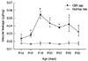 | Figure 1Time course of vascular permeability changes in the retina of OIR rats. Vascular permeability in the retina was measured at P14, P16, P18, P20, P22, P26 and P30. The Evans blue in the tissues was normalized by total protein concentration and expressed as µg of dye per mg of protein in the tissue. Values are mean ± SD (n = 7 to 10). (*p < 0.05: compared with OIR and control group) (OIR: Oxygen-induced retinopathy).
|
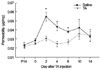 | Figure 2Time-dependent effects of TA on vascular hyperpermeability in the retinas of OIR rats. At P16 the right eye received an intravitreal injection of TA (4 mg per eye) and the left eye received saline. Vascular permeability was measured 2 and 0, 2, 4, 6, 10 and 14 days before and after the injection. The Evans blue in the tissues was normalized by total protein concentration and expressed as µg of dye per mg of protein in the tissue. Values are mean ± SD (n = 7 to 10). (*p < 0.05: compared with TA- and saline-treated group) (TA: triamcinolone acetonide).
|
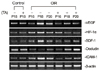 | Figure 3Expression of mRNAs analyzed by RT-PCR in the retina of both control and OIR rats and changes after TA treatment. This image is representative from six rats, showing PCR-amplified products of VEGF, HIF-1α, SDF-1, Occludin and ICAM-1, and β-actin as a loading control (TA: triamcinolone acetonide, OIR: oxygen-induced retinopathy).
|
References
1. Hebbandi SB, Bowen JR, Hipwell GC, et al. Ocular sequelae in extremely premature infants at 5 years of age. J Paediatr Child Health. 1997. 33:339–342.
2. Moore A. Retinopathy of prematurity. 1990. Boston: Blackwell Scientific;365–375.
3. Noma H, Funatsu H, Yamashita H, et al. Regulation of angiogenesis in diabetic retinopathy: possible balance between vascular endothelial growth factor and endostatin. Arch Ophthalmol. 2002. 120:1075–1080.
4. Raisler BJ, Berns KI, Grant MB, et al. Adeno-associated virus type-2 expression of pigmented epithelium-derived factor or Kringles 1-3 of angiostatin reduce retinal neovascularization. Proc Natl Acad Sci U S A. 2002. 99:8909–8914.
5. Adamis AP, Miller JW, Bernal MT, et al. Increased vascular endothelial growth factor levels in the vitreous of eyes with proliferative diabetic retinopathy. Am J Ophthalmol. 1994. 118:445–450.
6. Aiello LP, Avery RL, Arrigg PG, et al. Vascular endothelial growth factor in ocular fluid of patients with diabetic retinopathy and other retinal disorders. N Engl J Med. 1994. 331:1480–1487.
7. Malecaze F, Clamens S, Simorre-Pinatel V, et al. Detection of vascular endothelial growth factor messenger RNA and vascular endothelial growth factor-like activity in proliferative diabetic retinopathy. Arch Ophthalmol. 1994. 112:1476–1482.
8. Brafman A, Mett I, Shafir M, et al. Inhibition of oxygen-induced retinopathy in RTP801-deficient mice. Invest Ophthalmol Vis Sci. 2004. 45:3796–3805.
9. Treins C, Giorgetti-Peraldi S, Murdaca J, Van Obberghen E. Regulation of vascular endothelial growth factor expression by advanced glycation end products. J Biol Chem. 2001. 276:43836–43841.
10. Poulaki V, Qin W, Joussen AM, et al. Acute intensive insulin therapy exacerbates diabetic blood-retinal barrier breakdown via hypoxia-inducible factor-1alpha and VEGF. J Clin Invest. 2002. 109:805–815.
11. Grimm C, Wenzel A, Groszer M, et al. HIF-1-induced erythropoietin in the hypoxic retina protects against light-induced retinal degeneration. Nat Med. 2002. 8:718–724.
12. Grunewald M, Avraham I, Dor Y, et al. VEGF-induced adult neovascularization: recruitment, retention, and role of accessory cells. Cell. 2006. 124:175–189.
13. Lima e Silva R, Shen J, Hackett SF, et al. The SDF-1/CXCR4 ligand/receptor pair is an important contributor to several types of ocular neovascularization. FASEB J. 2007. 21:3219–3230.
14. Antonetti DA, Barber AJ, Khin S, et al. Penn State Retina Research Group. Vascular permeability in experimental diabetes is associated with reduced endothelial occludin content: vascular endothelial growth factor decreases occludin in retinal endothelial cells. Diabetes. 1998. 47:1953–1959.
15. McAllister IL, Vijayasekaran S, Chen SD, Yu DY. Effect of triamcinolone acetonide on vascular endothelial growth factor and occludin levels in branch retinal vein occlusion. Am J Ophthalmol. 2009. 147:838–846. 846.e1–846.e2.
16. Kim I, Moon SO, Kim SH, et al. Vascular endothelial growth factor expression of intercellular adhesion molecule 1 (ICAM-1), vascular cell adhesion molecule 1 (VCAM-1), and E-selectin through nuclear factor-kappa B activation in endothelial cells. J Biol Chem. 2001. 276:7614–7620.
17. Joussen AM, Poulaki V, Qin W, et al. Retinal vascular endothelial growth factor induces intercellular adhesion molecule-1 and endothelial nitric oxide synthase expression and initiates early diabetic retinal leukocyte adhesion in vivo. Am J Pathol. 2002. 160:501–509.
18. Clauss M, Gerlach M, Gerlach H, et al. Vascular permeability factor: a tumor-derived polypeptide that induces endothelial cell and monocyte procoagulant activity, and promotes monocyte migration. J Exp Med. 1990. 172:1535–1545.
19. Barleon B, Sozzani S, Zhou D, et al. Migration of human monocytes in response to vascular endothelial growth factor (VEGF) is mediated via the VEGF receptor flt-1. Blood. 1996. 87:3336–3343.
20. Sunderkötter C, Steinbrink K, Goebeler M, et al. Macrophages and angiogenesis. J Leukoc Biol. 1994. 55:410–422.
21. Ferrara N. Vascular endothelial growth factor: basic science and clinical progress. Endocr Rev. 2004. 25:581–611.
22. Sakurai E, Taguchi H, Anand A, et al. Targeted disruption of the CD18 or ICAM-1 gene inhibits choroidal neovascularization. Invest Ophthalmol Vis Sci. 2003. 44:2743–2749.
23. Tatar O, Adam A, Shinoda K, et al. Early effects of intravitreal triamcinolone acetonide on inflammation and proliferation in human choroidal neovascularization. Arch Ophthalmol. 2009. 127:275–281.
24. Kim YH, Chung IY, Choi MY, et al. Triamcinolone suppresses retinal vascular pathology via a potent interruption of proinflammatory signal-regulated activation of VEGF during a relative hypoxia. Neurobiol Dis. 2007. 26:569–576.
25. Antcliff RJ, Spalton DJ, Stanford MR, et al. Intravitreal triamcinolone for uveitic cystoid macular edema: an optical coherence tomography study. Ophthalmology. 2001. 108:765–772.
26. Young S, Larkin G, Branley M, Lightman S. Safety and efficacy of intravitreal triamcinolone for cystoid macular oedema in uveitis. Clin Experiment Ophthalmol. 2001. 29:2–6.
27. Jonas JB, Söfker A. Intraocular injection of crystalline cortisone as adjunctive treatment of diabetic macular edema. Am J Ophthalmol. 2001. 132:425–427.
28. Jonas JB, Hayler JK, Panda-Jonas S. Intravitreal injection of crystalline cortisone as adjunctive treatment of proliferative vitreoretinopathy. Br J Ophthalmol. 2000. 84:1064–1067.
29. Danis RP, Ciulla TA, Pratt LM, Anliker W. Intravitreal triamcinolone acetonide in exudative age-related macular degeneration. Retina. 2000. 20:244–250.
30. Challa JK, Gillies MC, Penfold PL, et al. Exudative macular degeneration and intravitreal triamcinolone: 18 month follow up. Aust N Z J Ophthalmol. 1998. 26:277–281.
31. Xu Q, Qaum T, Adamis AP. Sensitive blood-retinal barrier breakdown quantitation using Evans blue. Invest Ophthalmol Vis Sci. 2001. 42:789–794.
32. Kim YS, Kim YH, Cheon EW, et al. Retinal expression of clusterin in the streptozotocin-induced diabetic rat. Brain Res. 2003. 976:53–59.
33. Shih SC, Ju M, Liu N, Smith LE. Selective stimulation of VEGFR-1 prevents oxygen-induced retinal vascular degeneration in retinopathy of prematurity. J Clin Invest. 2003. 112:50–57.
34. Leske DA, Wu J, Fautsch MP, et al. The role of VEGF and IGF-1 in a hypercarbic oxygen-induced retinopathy rat model of ROP. Mol Vis. 2004. 10:43–50.
35. Zhang SX, Ma JX, Sima J, et al. Genetic difference in susceptibility to the blood-retina barrier breakdown in diabetes and oxygen-induced retinopathy. Am J Pathol. 2005. 166:313–321.
36. Gao G, Shao C, Zhang SX, et al. Kallikrein-binding protein inhibits retinal neovascularization and decreases vascular leakage. Diabetologia. 2003. 46:689–698.
37. Ozaki H, Hayashi H, Vinores SA, et al. Intravitreal sustained release of VEGF causes retinal neovascularization in rabbits and breakdown of the blood-retinal barrier in rabbits and primates. Exp Eye Res. 1997. 64:505–517.
38. Saishin Y, Saishin Y, Takahashi K, et al. VEGF-TRAP(R1R2) suppresses choroidal neovascularization and VEGF-induced breakdown of the blood-retinal barrier. J Cell Physiol. 2003. 195:241–248.
39. Nambu H, Nambu R, Oshima Y, et al. Angiopoietin 1 inhibits ocular neovascularization and breakdown of the blood-retinal barrier. Gene Ther. 2004. 11:865–873.
40. Houle JM, Strong HA. Duration of skin photosensitivity and incidence of photosensitivity reactions after administration of verteporfin. Retina. 2002. 22:691–697.
41. Brooks HL Jr, Caballero S Jr, Newell CK, et al. Vitreous levels of vascular endothelial growth factor and stromal-derived factor 1 in patients with diabetic retinopathy and cystoid macular edema before and after intraocular injection of triamcinolone. Arch Ophthalmol. 2004. 122:1801–1807.
42. Danis RP, Bingaman DP, Yang Y, Ladd B. Inhibition of preretinal and optic nerve head neovascularization in pigs by intravitreal triamcinolone acetonide. Ophthalmology. 1996. 103:2099–2104.
43. Jonas JB, Kreissig I, Kamppeter B, Degenring RF. [Intravitreal triamcinolone acetonide for the treatment of intraocular edematous and neovascular diseases]. Ophthalmologe. 2004. 101:113–120.
44. Lai DR, Chen HR, Lin LM, et al. Clinical evaluation of different treatment methods for oral submucous fibrosis. A 10-year experience with 150 cases. J Oral Pathol Med. 1995. 24:402–406.
45. Matsuda S, Gomi F, Oshima Y, et al. Vascular endothelial growth factor reduced and connective tissue growth factor induced by triamcinolone in ARPE19 cells under oxidative stress. Invest Ophthalmol Vis Sci. 2005. 46:1062–1068.
46. Lipworth BJ. Systemic adverse effects of inhaled corticosteroid therapy: A systematic review and meta-analysis. Arch Intern Med. 1999. 159:941–955.
47. Takahashi K, Saishin Y, Saishin Y, et al. Intraocular expression of endostatin reduces VEGF-induced retinal vascular permeability, neovascularization, and retinal detachment. FASEB J. 2003. 17:896–898.
48. Sima J, Zhang SX, Shao C, et al. The effect of angiostatin on vascular leakage and VEGF expression in rat retina. FEBS Lett. 2004. 564:19–23.
 XML Download