

 PDF
PDF ePub
ePub Citation
 Print
Print


Abstract
Purpose
To introduce a new surgical method of transscleral intraocular lens (IOL) fixation using a foldable, single-piece acrylic IOL with 4 loop haptics and to report the surgical results.
Methods
After a single-piece acrylic IOL with 4 loop haptics was injected into the anterior chamber and positioned on top of the iris diaphragm, a 10-0 Prolene STC-6 straight needle and a 27-gauge needle were used to string the prolene thread through the haptic openings from front to back fixating the IOL to the sclera, resulting in a transscleral "1 loop 4 points" fixation. Twenty-eight eyes of 28 patients who had received transscleral fixation via this new technique were retrospectively reviewed. The best corrected vision acuity (BCVA) was measured after a postoperative period of at least 6 months. Intraoperative and postoperative complications were investigated.
Results
In 27 out of 28 eyes (96.4%), the postoperative BCVA was better than 0.5 (Snellen chart). The only complication found was 1 case of choroidal detachment (3.6%).
Conclusions
The new transscleral "1 loop 4 points" fixation technique of a foldable, single-piece acrylic IOL in the absence of capsular support is an easy procedure and reduces surgical time and hastens visual rehabilitation due to excellent IOL positioning stability. Additionally, the technique described in the present study may be a safe procedure with minimal complications.
Figures and Tables
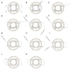 | Figure 1(A) The intraocular lens (IOL) is positioned on top of the iris diaphragm. The entry sites A,B,C, and D are marked 2.0 mm from the limbus and 6.0 mm from each other. (B) After using a hook to push one of the haptics behind the iris diaphragm, a double armed 10-0 prolene STC-6 straight needle is threaded through the haptic loop from front to back. (C) A 27-gauge needle is used to enter the sclera at site A and threaded through the haptic loop in the same manner as the prolene needle. (D, E) The prolene needle is docked within the 27 gauge needle and pulled out of the opposite sclera site A. (F-I) The second prolene needle is entered the sclera at site D and retrieved within the 27-gauge needle in the same manner as the first needle. (J) The sutures are tightened 3 times using a square knot. (K) The knot is rotated and buried into the scleral through site A.
|
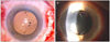 | Figure 2(A) Immediate postoperative photograph shows the intraocular lens (IOL) was positioned correctly. The 2 black arrows indicate the prolene sutures which are located behind the IOL optic. (B) Postoperative photograph 1 year after undergoing '1 loop - 4 points' shows no definite tilting or decentering of the IOL. The 2 white arrows also indicate the prolene sutures.
|
References
1. Güell JL, Barrera A, Manero F. A review of suturing techniques for posterior chamber lenses. Curr Opin Ophthalmol. 2004. 15:44–50.
2. Oh HS, Chu YK, Kwon OW. Surgical technique for suture fixation of a single-piece hydrophilic acrylic intraocular lens in the absence of capsule support. J Cataract Refract Surg. 2007. 33:962–965.
3. Lewis JS. Ab externo sulcus fixation. Ophthalmic Surg. 1991. 22:692–695.
4. Michaeli A, Assia EI. Scleral and iris fixation of posterior chamber lenses in the absence of capsular support. Curr Opin Ophthalmol. 2005. 16:57–60.
5. Por YM, Lavin MJ. Techniques of intraocular lens suspension in the absence of capsular/zonular support. Surv Ophthalmol. 2005. 50:429–462.
6. Wagoner MD, Cox TA, Ariyasu RG, et al. Intraocular lens implantation in the absence of capsular support: a report by the American Academy of Ophthalmology. Ophthalmology. 2003. 110:840–859.
7. Yang JY, Ma KT, Kim JH. Choice of one-piece intraocular lens power and changes of anterior chamber in sulcus implantation due to posterior capsular rupture during Cataract Surgery. J Korean Ophthalmol Soc. 2012. 53:775–780.
8. Duffey RJ, Holland EJ, Agapitos PJ, Lindstrom RL. Anatomic study of transsclerally sutured intraocular lens implantation. Am J Ophthalmol. 1989. 108:300–309.
9. Althaus C, Sundmacher R. [Endoscopically controlled optimization of trans-scleral suture fixation of posterior chamber lenses in the ciliary sulcus]. Ophthalmologe. 1993. 90:317–324.
10. Bellucci R, Pucci V, Morselli S, Bonomi L. Secondary implantation of angle-supported anterior chamber and scleral-fixated posterior chamber intraocular lenses. J Cataract Refract Surg. 1996. 22:247–252.
11. Tabandeh H, Flynn HW Jr. Suprachoroidal hemorrhage during pars plana vitrectomy. Curr Opin Ophthalmol. 2001. 12:179–185.
12. Price FW Jr, Whitson WE. Suprachoroidal hemorrhage after placement of a scleral-fixated lens. J Cataract Refract Surg. 1990. 16:514–515.
 XML Download