

 PDF
PDF ePub
ePub Citation
 Print
Print


Abstract
Purpose
To introduce a more efficient and time-saving scleral fixation technique for a posterior chamber foldable intraocular lens and to report the clinical results.
Methods
A foldable acrylic 3-Piece IOL was sutured to the sclera via a small corneal incision. The guiding hollow needle was not used, which differs from other ab externo techniques. Instead, the curved long needle was directly pulled out through the cornea. The scleral flap was not used to bury the scleral suture knot; Instead, the scleral suture knot was translocated at the temporal area.
Figures and Tables
Figure 1
Summary of the Surgical procedure. (A) After careful conjunctival periotomy, two entry sites directed at 5 and 11 o'clock (point A and B) were marked on the sclera, 1.0 mm posterior to the limbus. (B) Two long curved needles with a double-armed 10-0 polypropylene suture were used in this procedure. (C) Different from other ab externo techniques, the needle was pulled out directly through the peripheral cornea (the yellow circle). The green arrow indicates direction of the needle. (D) The second needle passed through the sclera at the 180 degree opposite direction of the first entry site (point B). (E) The sutures were tied securely to each haptic of 3-piece foldable acrylic IOL. (F) The long curved needle was passed from each point of fixation through the sclera, 3 mm posterior and parallel to the limbus. Each intra-scleral suture is advanced in a lamellar fashion and directed to the temporal area to avoid exposure which can be promoted by frictional blinking movement. The schematic drawing shows how the suture is placed. Two red dot lines in the schematic drawing show the passage of the sutures. The green arrow in the raw figure and the red star in the schematic drawing indicate the suture knot left directly over the bare sclera without burying it.
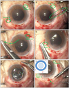
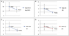
Figure 2
Mean BCVA and refractive index, Mean BCVA (log MAR), SE (D) and Sph (D) showed statistically significant differences after surgery (p < 0.001, p = 0.002, p = 0.005, respectively). Only Cyl value showed statistically insignificant result. BCVA = best corrected visual acuity; SE = spherical equivalent; Sph = spherical refraction; Cyl = cylindrical refraction.


References
1. Malbran ES, Malbran E Jr, Negri I. Lens guide suture for transport and fixation in secondary IOL implantation after intracapsular extraction. Int Ophthalmol. 1986. 9:151–160.
2. Regillo CD, Tidwell J. A small-incision technique for suturing a posterior chamber intraocular lens. Ophthalmic Surg Lasers. 1996. 27:473–475.
3. Kaynak S, Ozbek Z, Pasa E, et al. Transscleral fixation of foldable intraocular lenses. J Cataract Refract Surg. 2004. 30:854–857.
4. Yepez JB, de Yepez JC, Valero A, Arevalo JF. Surgical technique for transscleral fixation of a foldable posterior chamber intraocular lens. Ophthalmic Surg Lasers Imaging. 2006. 37:247–250.
5. Taskapili M, Gulkilik G, Engin G, et al. Transscleral fixation of a single-piece hydrophilic foldable acrylic intraocular lens. Can J Ophthalmol. 2007. 42:256–261.
6. Oh HS, Chu YK, Kwon OW. Surgical technique for suture fixation of a single-piece hydrophilic acrylic intraocular lens in the absence of capsule support. J Cataract Refract Surg. 2007. 33:962–965.
7. Szurman P, Petermeier K, Jaissle GB, Bartz-Schmidt KU. A new small-incision technique for injector implantation of transsclerally sutured foldable lenses. Ophthalmic Surg Lasers Imaging. 2007. 38:76–80.
8. Fass ON, Herman WK. Sutured intraocular lens placement in aphakic post-vitrectomy eyes via small-incision surgery. J Cataract Refract Surg. 2009. 35:1492–1497.
9. Kim SJ, Lee SJ, Park CH, et al. Long-term stability and visual outcomes of a single-piece, foldable, acrylic intraocular lens for scleral fixation. Retina. 2009. 29:91–97.
10. Taskapili M, Gulkilik G, Kocabora MS, Nilay K. Transscleral fixation of single-piece hydrophilic acrylic lenses with no eyelets. Ophthalmic Surg Lasers Imaging. 2009. 40:434–436.
11. Yaguchi S, Yaguchi S, Noda Y, et al. Foldable acrylic intraocular lens with distended haptics for transscleral fixation. J Cataract Refract Surg. 2009. 35:2047–2050.
12. Schechter RJ. Suture-wick endophthalmitis with sutured posterior chamber intraocular lenses. J Cataract Refract Surg. 1990. 16:755–756.
13. Lewis JS. Ab externo sulcus fixation. Ophthalmic Surg. 1991. 22:692–695.
14. Choi KS, Park SY, Sun HJ. Transscleral fixation by injector implantation of a foldable intraocular lens. Ophthalmic Surg Lasers Imaging. 2010. 41:272–275.
15. Kim DH, Heo JW, Hwang SW, et al. Modified transscleral fixation using combined temporary haptic externalization and injector intraocular lens implantation. J Cataract Refract Surg. 2010. 36:707–711.
16. Lewis JS. Sulcus fixation without flaps. Ophthalmology. 1993. 100:1346–1350.
17. Baykara M, Avci R. Prevention of suture knot exposure in posterior chamber intraocular lens implantation by 4-point scleral fixation technique. Ophthalmic Surg Lasers Imaging. 2004. 35:379–382.
18. Hoffman RS, Fine IH, Packer M. Scleral fixation without conjunctival dissection. J Cataract Refract Surg. 2006. 32:1907–1912.
19. Szurman P, Petermeier K, Aisenbrey S, et al. Z-suture: a new knotless technique for transscleral suture fixation of intraocular implants. Br J Ophthalmol. 2010. 94:167–169.
20. Solomon K, Gussler JR, Gussler C, Van Meter WS. Incidence and management of complications of transsclerally sutured posterior chamber lenses. J Cataract Refract Surg. 1993. 19:488–493.
 XML Download