

 PDF
PDF ePub
ePub Citation
 Print
Print


Abstract
Purpose
To evaluate the parameters affecting vaulting and correlation between preoperative crystalline lens rise and vaulting after implantable collamer lense (ICL) implantation.
Methods
A total of 53 eyes of 34 patients who underwent ICL implantation were examined retrospectively. White-to-white (WTW) and anterior chamber depth (ACD) were obtained from scanning topography (ORB scan) before surgery. Preoperative crystalline lens rise (CLR) and vaulting at 6 months after ICL implantation were measured using anterior segment optic coherence tomography (AS-OCT). Multiple regression analysis was performed to evaluate the factors affecting central vaulting.
Results
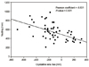
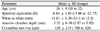
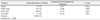
The mean preoperative crystalline lens rise was -120 ± 219 µm, and mean central vaulting 6 months after surgery was 544 ± 175 µm. Preoperative SE, WTW, ACD, and CLR were significantly correlated with vaulting at 6 months after surgery. With the use of meaningful variables, multiple regression analysis showed that CLR, WTW, ACD and SE, in that order of influence, had significant effects on vaulting and the multiple regression equation was obtained as follows: Vaulting (µm) = (160.913 × ACD (mm)) + (170.134 × WTW (mm)) + (-0.338 × CLR (µm)) + (-23.783 × SE (D)) - 2250.184.
Figures and Tables
Figure 1
Crystalline lens rise which is the distance between the anterior pole of the crystalline lens and the line connecting the 3 o'clock to 9 o'clock angle recess is measured on the horizontal diameter median image of anterior segment OCT. In this patient, crystalline lens rise is 320 µm.
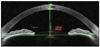
Figure 2
Anterior segment OCT image of the left eye of a 25-year-old man at 6 months after ICL implantation. Vaulting is 410 µm.
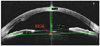
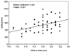
Figure 4
Correlation between horizontal white-to-white distance and the central vaulting of the ICL.


References
1. Uusitalo RJ, Aine E, Sen NH, Laatikainen L. Implantable contact lens for high myopia. J Cataract Refract Surg. 2002. 28:29–36.
2. Sanders DR, Vukich JA, Doney K, Gaston M. Implantable Contact Lens in Treatment of Myopia Study Group. U.S. Food and Drug Administration clinical trial of the implantable Contact Lens for moderate to high myopia. Ophthalmology. 2003. 110:255–266.
3. Arne JL, Lesueur LC. Phakic posterior chamber lenses for high myopia: functional and anatomical outcomes. J Cataract Refract Surg. 2000. 26:369–374.
4. Zaldivar R, Davidorf JM, Oscherow S. Posterior chamber phakic intraocular lens for myopia of -8 to -19 diopters. J Refract Surg. 1998. 14:294–305.
5. Chun YS, Lee JH, Lee JM, et al. Outcomes after implantable contact lens for moderate to high myopia. J Korean Ophthalmol Soc. 2004. 45:480–489.
6. Dejaco-Ruhswurm I, Scholz U, Pieh S, et al. Long-term endothelial changes in phakic eyes with posterior chamber intraocular lenses. J Cataract Refract Surg. 2002. 28:1589–1593.
7. Jiménez-Alfaro I, Gómez-Tellería G, Bueno JL, Puy P. Contrast sensitivity after posterior chamber phakic intraocular lens implantation for high myopia. J Refract Surg. 2001. 17:641–645.
8. Lee SY, Cheon HJ, Baek TM, Lee KH. Implantable contact lens to correct high myopia (clinical study with 24 months follow-up). J Korean Ophthalmol Soc. 2000. 41:1515–1522.
9. Chun YS, Lee JH, Lee JM, Park IK. IOP and gonioscopic changes after implantable contact lens implantation in myopic eyes. J Korean Ophthalmol Soc. 2005. 46:336–344.
10. Kamiya K, Shimizu K, Komatsu M. Factors affecting vaulting after implantable collamer lens implantation. J Refract Surg. 2009. 25:259–264.
11. Lee DH, Choi SH, Chung ES, Chung TY. Correlation between Preoperative biometry and posterior chamber phakic Visian Implantable Collamer Lens Vaulting. Ophthalmology. 2012. 119:272–277.
12. Baïkoff G, Bourgeon G, Jodai HJ, et al. Pigment dispersion and Artisan phakic intraocular lenses: crystalline lens rise as a safety criterion. J Cataract Refract Surg. 2005. 31:674–680.
13. Baïkoff G. Anterior segment OCT and phakic intraocular lenses: a perspective. J Cataract Refract Surg. 2006. 32:1827–1835.
14. Kucumen RB, Yenerel NM, Gorgun E, et al. Anterior segment optical coherence tomography measurement of anterior chamber depth and angle changes after phacoemulsification and intraocular lens implantation. J Cataract Refract Surg. 2008. 34:1694–1698.
15. Lackner B, Pieh S, Schmidinger G, et al. Long-term results of implantation of phakic posterior chamber intraocular lenses. J Cataract Refract Surg. 2004. 30:2269–2276.
16. Bourne WM, Nelson LR, Buller CR, et al. Long-term observation of morphologic and functional features of cat corneal endothelium after wounding. Invest Ophthalmol Vis Sci. 1994. 35:891–899.
17. Güell JL, Morral M, Gris O, et al. Evaluation of Verisyse and Artiflex phakic intraocular lenses during accommodation using Visante optical coherence tomography. J Cataract Refract Surg. 2007. 33:1398–1404.
18. Bechmann M, Ullrich S, Thiel MJ, et al. Imaging of posterior chamber phakic intraocular lens by optical coherence tomography. J Cataract Refract Surg. 2002. 28:360–363.
19. Petternel V, Köppl CM, Dejaco-Ruhswurm I, et al. Effect of accommodation and pupil size on the movement of a posterior chamber lens in the phakic eye. Ophthalmology. 2004. 111:325–331.
20. García-Feijoó J, Alfaro IJ, Cuiña-Sardiña R, et al. Ultrasound biomicroscopy examination of posterior chamber phakic intraocular lens position. Ophthalmology. 2003. 110:163–172.
21. Gonvers M, Bornet C, Othenin-Girard P. Implantable contact lens for moderate to high myopia: relationship of vaulting to cataract formation. J Cataract Refract Surg. 2003. 29:918–924.
22. Schmidinger G, Lackner B, Pieh S, Skorpik C. Long-term changes in posterior chamber phakic intraocular collamer lens vaulting in myopic patients. Ophthalmology. 2010. 117:1506–1511.
 XML Download