

 PDF
PDF ePub
ePub Citation
 Print
Print


Abstract
Purpose
The present study assessed the diagnostic significance of digital infrared thermal imaging (DITI) in determining the inflammatory state of patients with nonspecific orbital inflammation.
Methods
The present study included 11 patients diagnosed with nonspecific orbital inflammation between December 2009 and March 2011 and who were followed-up for more than 6 months. All patients were all diagnosed based on computed tomography (CT), magnetic resonance image (MRI) and tissue biopsy. The grade of eyelid swelling was classified as 5 and severe compared with the temperature of location. The temperature of the upper eyelid, caruncle, medial conjunctiva, lateral conjunctiva, lower eyelid, cornea and lateral orbit were measured with DITI.
Results
When comparing the normal eye with the diseased eye in patients with nonspecific orbital inflammation, the temperature of the upper eyelid and cornea were statistically significant (p = 0.003, p = 0.038, respectively, Mann-Whitney test). The correlation between the grade of eyelid swelling and the temperature of location was most highly related in the temperature of the upper eyelid (Spearman's correlation coefficient, r = 0.55, p = 0.008) and cornea (Spearman's correlation coefficient, r = 0.45, p = 0.037).
Conclusions
DITI may aid in evaluating the inflammatory state of nonspecific orbital inflammation. In particular, the temperature of the upper eyelid and cornea can be very useful indicator. Future studies, including larger study population are necessary in order to confirm DITI as a diagnostic tool which can assess the results of medical treatment by comparing temperature before and after treatment.
Figures and Tables
Figure 1
Examples of eyelid swelling by severity grade. (A) normal eye (Grade 1), (B) mild swelling (Grade 2): apical lesion, (C) moderate swelling (Grade 3): preseptal lesion, (D) severe swelling (Grade 4): lacrimal gland lesion, (E) very severe swelling (Grade 5): diffuse lesion (lowerlid margin involved nearly pupil center due to upperlid swelling).
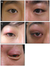
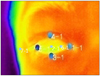
Figure 2
The exact point of right lateral orbit which were measured by DITI. 1-1: caruncle, 2-1: cornea, 3-1: lower eyelid, 4-1: upper eyelid, 5-1: lateral conjunctiva, 6-1: medial conjunctiva, 7-1: lateral orbit (There is no photographic image with all seven points demarcated, so 2-1, 6-1 and 7-1 were marked with animation to explain exact point).

References
1. Park SJ, Sin SJ, Lee DG, Jang JW. Pseudotumor: distribution, clinical features, treatment outcomes. J Korean Ophthalmol Soc. 2008. 49:1379–1386.
2. Lee H, Kim SJ, Lee SY. Classification and treatment efficacy of orbital pseudotumor. J Korean Ophthalmol Soc. 2001. 42:1647–1654.
3. Caminha LS, Pinto ER, Sousa PA, et al. Orbital pseudotumor: a differential diagnosis of Graves' ophthalmopathy. Arq Bras Endocrinol Metabol. 2011. 55:85–88.
4. Jones BF. A reappraisal of the use of infrared thermal image analysis in medicine. IEEE Trans Med Imaging. 1998. 17:1019–1027.
5. Jiang LJ, Ng EY, Yeo AC, et al. A perspective on medical infrared imaging. J Med Eng Technol. 2005. 29:257–267.
6. Sun PC, Lin HD, Jao SH, et al. Relationship of skin temperature to sympathetic dysfunction in diabetic at-risk feet. Diabetes Res Clin Pract. 2006. 73:41–46.
7. Singh G, Bhinder HS. Comparison of noncontact infrared and remote sensor thermometry in normal and dry eye patients. Eur J Ophthalmol. 2005. 15:668–673.
8. Chang TC, Hsiao YL, Liao SL. Application of digital infrared thermal imaging in determining inflammatory state and follow-up effect of methylprednisolone pulse therapy in patients with Graves' ophthalmopathy. Graefes Arch Clin Exp Ophthalmol. 2008. 246:45–49.
9. Shih SR, Li HY, Hsiao YL, Chang TC. The application of temperature measurement of the eyes by digital infrared thermal imaging as a prognostic factor of methylprednisolone pulse therapy for Graves' ophthalmopathy. Acta Ophthalmol. 2010. 88:e154–e159.
10. Niehof SP, Huygen FJ, van der Weerd RW, et al. Thermography imaging during static and controlled thermoregulation in complex regional pain syndrome type 1: diagnostic value and involvement of the central sympathetic system. Biomed Eng Online. 2006. 5:30.
11. Zaproudina N, Ming Z, Hänninen OO. Plantar infrared thermography measurements and low back pain intensity. J Manipulative Physiol Ther. 2006. 29:219–223.
12. Kontos M, Wilson R, Fentiman I. Digital infrared thermal imaging (DITI) of breast lesions: sensitivity and specificity of detection of primary breast cancers. Clin Radiol. 2011. 66:536–539.
 XML Download