

 PDF
PDF ePub
ePub Citation
 Print
Print


Abstract
Purpose
To report a case of bilateral retinal detachment in a 15-year-old child with Tourette syndrome.
Case summary
A 15-year-old child treated for Tourette syndrome for 4 years presented with decreased visual acuity of several days in duration. The fundus of the right eye was not observed due to lens opacity and posterior synechiae. The B-scan of the right eye showed funnel-shaped densely reflective echoes connected to the optic disc, suggesting a total retinal detachment. Fundus examination of the left eye revealed an inverted retinal flap, which covered the posterior pole. During vitrectomy of the left eye, a ciliary body detachment anterior to a giant retinal tear extending 360 degrees was observed. In addition, an inverted flap covering 2 superior retinal quadrants was observed. A perfluorocarbon liquid was injected to unfold the tear's inverted flap, and silicone oil tamponade was performed.
Conclusions
Self-induced and repeated periocular trauma induced by motor tics of Tourette syndrome can result in bilateral retinal detachment. Regular ophthalmic examinations are helpful for early detection of ocular complications including periocular trauma induced by motor tics of Tourette syndrome.
Figures and Tables
Figure 1
(A) Slit lamp biomicroscopy of the right eye showed whitish lens opacity and posterior synechiae. (B) B-scan revealed the funnel-shaped densely reflective echoes, which suggested total retinal detachment connected to the optic disc.
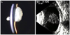
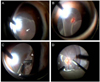
Figure 2
Photographs showing fundus findings captured during vitrectomy. (A) The surgeon could see the optic disc (white arrow) beneath the inverted retinal flap. (B) After lifting the flap, clear optic disc with retinal vessels were seen. (C) During peripheral vitrectomy with indentation, ciliary body detachment (arrow head) anterior to retinal tear (white arrow) was seen. (D) The perfluorocarbon liquid was injected to unfold the inverted flap of the tear.
References
1. Jankovic J. Tourette's syndrome. N Engl J Med. 2001. 345:1184–1192.
2. Lim S, Rezai KA, Abrams GW, Eliott D. Self-induced, bilateral retinal detachment in Tourette syndrome. Arch Ophthalmol. 2004. 122:930–931.
3. Du JC, Chiu TF, Lee KM, et al. Tourette syndrome in children: an updated review. Pediatr Neonatol. 2010. 51:255–264.
4. American Psychiatric Association. Diagnostic and Statistical Manual of Mental Disorders. 1994. 4th ed. Washington DC: American Psychiatric Association;100–105.
5. Tatlipinar S, Iener EC, Ilhan B, Semerci B. Ophthalmic manifestations of Gilles de la Tourette syndrome. Eur J Ophthalmol. 2001. 11:223–226.
6. Jung HY, Chung SJ, Hwang JM. Tic disorders in children with frequent eye blinking. J AAPOS. 2004. 8:171–174.
7. Margo CE. Tourette syndrome and iatrogenic eye injury. Am J Ophthalmol. 2002. 134:784–785.
8. Robertson MM, Trimble MR, Lees AJ. Self-injurious behaviour and the Gilles de la Tourette syndrome: a clinical study and review of the literature. Psychol Med. 1989. 19:611–625.
9. Kandarakis A, Karampelas M, Soumplis V, et al. A case of bilateral self-induced keratoconus in a patient with tourette syndrome associated with compulsive eye rubbing: case report. BMC Ophthalmol. 2011. 11:28.
10. Mashor RS, Kumar NL, Ritenour RJ, Rootman DS. Keratoconus caused by eye rubbing in patients with Tourette Syndrome. Can J Ophthalmol. 2011. 46:83–86.
11. Goffstein R, Burton TC. Differentiating traumatic from nontraumatic retinal detachment. Ophthalmology. 1982. 89:361–368.
 XML Download