

 PDF
PDF ePub
ePub Citation
 Print
Print


Abstract
Purpose
To report 2 young female patients with bilateral posterior scleritis and serous retinal detachment.
Case summary
An 11-year-old girl (Case 1) and a 16-year-old girl (Case 2) visited our clinic with bilateral ocular pain, redness, and blurred vision. Slit lamp examinations revealed severe bilateral scleral injection and mild anterior chamber reactions. Fundus examinations showed bilateral serous retinal detachments in the macular area. In both patients, diffuse multifocal leaking and pooling were found at the macula in the early and late phase fluorescein angiography, respectively. On the B-mode ultrasounds and orbital images (MRI or CT), scleral thickening with retention of subtenons fluid were found. There were no systemic diseases associated with the conditions. We diagnosed the patients with bilateral posterior scleritis and administered systemic steroids. After systemic steroid treatment, all of the symptoms were alleviated. Three months after the regression, bilateral posterior scleritis recurred in patient 2. Oral cyclosporine 100 mg was additionally prescribed in addition to the previous medications that she had taken during her first treatment.
Figures and Tables
Figure 1
(Case 1) External photograph demonstrates conjunctival and scleral injections. There was no restriction of eyeball movements.
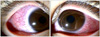
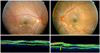
Figure 2
(Case 1) Fundus photographs and optical coherence tomography taken at the time of initial examinations show broad serous retinal detachment in the macular area.
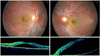
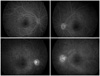
Figure 3
(Case 1) Early fluorescein angiography shows diffuse leakage of dye. Late fluorescein angiography shows pooling of fluorescein at the serous retinal detachment area and optic disc leakage.
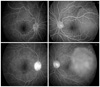
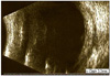
Figure 4
(Case 1) Ultrasound scan of the left eye (Case 1) shows diffuse scleral and choroidal thickening (2.2 mm) and presence of edema in the Tenon's space (arrow).
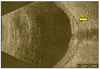
Figure 5
(Case 1) Orbital computed tomograp- hic scan shows scleral thickening (arrow) in both eyes, especially in the left side.
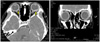
Figure 6
(Case 2) Fundus photographs taken at the time of initial examinations show serous retinal detachments in both sides and hyperemia of the left optic disc. OCT scan shows serous retinal detachments in both sides and cyst-like formations underneath the retina (arrow) in the left eye.
References
1. McCluskey PJ, Watson PG, Lightman S, et al. Posterior scleritis: clinical features, systemic associations, and outcome in a large series of patients. Ophthalmology. 1999. 106:2380–2386.
2. Benson WE. Posterior scleritis. Surv Ophthalmol. 1988. 32:297–316.
3. Wald KJ, Spaide R, Patalano VJ, et al. Posterior scleritis in children. Am J Ophthalmol. 1992. 113:281–286.
4. Tsujikawa A, Yamashiro K, Yamamoto K, et al. Retinal cystoid spaces in acute Vogt-Koyanagi-Harada syndrome. Am J Ophthalmol. 2005. 139:670–677.
5. Erdol H, Kola M, Turk A. Optical coherence tomography findings in a child with posterior scleritis. Eur J Ophthalmol. 2008. 18:1007–1010.
6. Jensen JE, Fledelius HC, Prause JU, Scherfig E. An unusual ophthalmic tumour in a 5-year-old boy. Acta Ophthalmol Suppl. 1992. 204:110–102.
7. Foster CS, de la Maza M Sa. The sclera. 1994. New York: Springer-Verlag;112–123.
8. Woon WH, Stanford MR, Graham EM. Severe idiopathic posterior scleritis in children. Eye. 1995. 9:570–574.
9. Watson PG, Young RD. Scleral structure, organisation and disease. A review. Exp Eye Res. 2004. 78:609–623.
10. McCluskey P, Wakefield D. Intravenous pulse methylprednisolone in scleritis. Arch Ophthalmol. 1987. 105:793–797.
11. Wakefield D, McCluskey P. Cyclosporin therapy for severe scleritis. Br J Ophthalmol. 1989. 73:743–746.
12. Horo S, Sudharshan S, Biswas J. Recurrent posterior scleritis-report of a case. Ocul Immunol Inflamm. 2006. 14:51–56.
 XML Download