

 PDF
PDF ePub
ePub Citation
 Print
Print


Abstract
Purpose
To compare the corneal endothelial cell changes in both eyes of Korean patients with clinically unilateral exfoliation syndrome using specular microscopy.
Methods
A total of 144 eyes of 72 patients diagnosed with clinically unilateral exfoliation syndrome at Yeungnam University Hospital between March 2000 and February 2011 were retrospectively reviewed. Comparisons of corneal morphometric analysis were made including endothelial cell density, coefficient of variation, hexagonality, and central corneal thickness between the exfoliative and fellow non-exfoliative eyes in 72 patients with naive unilateral exfoliation syndrome. If patients received intraocular surgery during the follow-up periods, the number of intraocular surgeries and changes of the above-mentioned morphometric analysis were evaluated.
Results
The paired exfoliative and fellow non-exfoliative eyes did not differ in endothelial cell density (2587.0 ± 391.0 vs. 2626.8 ± 354.6 cells/mm2, p = 0.321), in the coefficient of variation of cell size (35.9 ± 5.1 vs. 37.1 ± 4.7%), hexagonality (59.5 ± 7.3 vs. 57.8 ± 6.3%), and central corneal thickness (530.5 ± 37.6 vs. 532.0 ± 35.2 µm). However, the exfoliative eyes had significantly higher values for the number of intraocular surgeries (0.97 ± 0.78 vs. 0.28 ± 0.48, p < 0.001) and decrement of corneal endothelial cells (410.9 ± 538.7 vs. 19.0 ± 284.5 cells/mm2, p = 0.007).
Conclusions
There were no significant morphologic differences in corneal endothelium between exfoliative eyes and fellow eyes in the present study. However, the authors suggest that specular microscopic examination be performed before intraocular surgery in eyes with exfoliation syndrome when considering the higher frequency of intraocular surgeries and the resultant corneal endothelial damages observed in the present study.
Figures and Tables
Table 2
Findings in specular microscopy, pachymetry and number of intraocular surgery
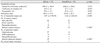
Values are presented as mean ± SD unless otherwise indicated.
*p-value by paired t-test; †p-value by Pearson chi-square test.
XFS = exfoliative eye; Non-XFS = fellow (non-exfoliative) eye; PEA = phacoemulsification; PCL = posterior chamber lens implantation; ECCE = extracapsular cataract extraction; GDD = glaucoma drainage device.


References
1. Roth M, Epstein DL. Exfoliation syndrome. Am J Ophthalmol. 1980. 89:477–481.
2. Naumann GO, Schlötzer-Schrehardt U, Küchle M. Pseudoexfoliation syndrome for the comprehensive ophthalmologist. Intraocular and systemic manifestations. Ophthalmology. 1998. 105:951–968.
3. Ritch R, Schlötzer-Schrehardt U. Exfoliation syndrome. Surv Ophthalmol. 2001. 45:265–315.
4. Schlötzer-Schrehardt U, Naumann GO. Ocular and systemic pseudoexfoliation syndrome. Am J Ophthalmol. 2006. 141:921–937.
5. Ritch R. Exfoliation syndrome-the most common identifiable cause of open-angle glaucoma. J Glaucoma. 1994. 3:176–177.
6. Ritch R. Exfoliation syndrome. Curr Opin Ophthalmol. 2001. 12:124–130.
7. Futa R, Shimizu T, Furuyoshi N, et al. Clinical features of capsular glaucoma in comparison with primary open-angle glaucoma in Japan. Acta Ophthalmol (Copenh). 1992. 70:214–219.
8. Aasved H. Prevalence of fibrillopathia epitheliocapsularis (pseudoexfoliation) and capsular glaucoma. Trans Ophthalmol Soc U K. 1979. 99:293–295.
9. Cho SW, Kim JM, Choi CY, Park KH. Changes in corneal endothelial cell density in patients with normal-tension glaucoma. Jpn J Ophthalmol. 2009. 53:569–573.
10. Hau S, Barton K. Corneal complications of glaucoma surgery. Curr Opin Ophthalmol. 2009. 20:131–136.
11. Gharagozloo NZ, Baker RH, Brubaker RF. Aqueous dynamics in exfoliation syndrome. Am J Ophthalmol. 1992. 114:473–478.
12. Tanhehco T, Chen SH. Pseudoexfoliation syndrome and cataract surgery. Int Ophthalmol Clin. 2010. 50:81–93.
13. Ishikawa A. Risk factors for reduced corneal endothelial cell density before cataract surgery. J Cataract Refract Surg. 2002. 28:1982–1992.
14. Miyake K, Matsuda M, Inaba M. Corneal endothelial changes in pseudoexfoliation syndrome. Am J Ophthalmol. 1989. 108:49–52.
15. Naumann GO, Küchle M. Primary corneal graft failure. Arch Ophthalmol. 1996. 114:1031.
16. Naumann GO, Schlötzer-Schrehardt U. Keratopathy in pseudoexfoliation syndrome as a cause of corneal endothelial decompensation: a clinicopathologic study. Ophthalmology. 2000. 107:1111–1124.
17. Quiroga L, Lansingh VC, Samudio M, et al. Characteristics of the corneal endothelium and pseudoexfoliation syndrome in patients with senile cataract. Clin Experiment Ophthalmol. 2010. 38:449–455.
18. Zheng X, Shiraishi A, Okuma S, et al. In vivo confocal microscopic evidence of keratopathy in patients with pseudoexfoliation syndrome. Invest Ophthalmol Vis Sci. 2011. 52:1755–1761.
19. Ringvold A. Corneal endothelial involvement in pseudoexfoliation syndrome. Arch Ophthalmol. 1994. 112:297–298.
20. Wirbelauer C, Anders N, Pham DT, Wollensak J. Corneal endothelial cell changes in pseudoexfoliation syndrome after cataract surgery. Arch Ophthalmol. 1998. 116:145–149.
21. Ehlers N, Hansen FK, Aasved H. Biometric correlations of corneal thickness. Acta Ophthalmol (Copenh). 1975. 53:652–659.
22. Puska P, Vasara K, Harju M, Setälä K. Corneal thickness and corneal endothelium in normotensive subjects with unilateral exfoliation syndrome. Graefes Arch Clin Exp Ophthalmol. 2000. 238:659–663.
23. Chang WH, Cha SC. Clinical manifestations of exfoliation syndrome in Korea. J Korean Ophthalmol Soc. 2000. 41:1768–1774.
24. Choi J, Park KH. Clinical characteristics of Korean patients with pseudoexfoliation. J Korean Ophthalmol Soc. 2006. 47:577–586.
25. Stefaniotou M, Kalogeropoulos C, Razis N, Psilas K. The cornea in exfoliation syndrome. Doc Ophthalmol. 1992. 80:329–333.
26. Schlötzer-Schrehardt UM, Dörfler S, Naumann GO. Corneal endothelial involvement in pseudoexfoliation syndrome. Arch Ophthalmol. 1993. 111:666–674.
27. Vesti E, Kivelä T. Exfoliation syndrome and exfoliation glaucoma. Prog Retin Eye Res. 2000. 19:345–368.
28. Tarkkanen A, Kivelä T. Cumulative incidence of converting from clinically unilateral to bilateral exfoliation syndrome. J Glaucoma. 2004. 13:181–184.
29. Kivelä T, Hietanen J, Uusitalo M. Autopsy analysis of clinically unilateral exfoliation syndrome. Invest Ophthalmol Vis Sci. 1997. 38:2008–2015.
30. Hammer T, Schlötzer-Schrehardt U, Naumann GO. Unilateral or asymmetric pseudoexfoliation syndrome? An ultrastructural study. Arch Ophthalmol. 2001. 119:1023–1031.
31. Kim KS, Park SY, Oh JS. Morphometric analysis of the corneal endothelial cells in normal Korean. J Korean Ophthalmol Soc. 1992. 33:320–325.
32. Laatikainen L. Fluorescein angiographic studies of the peripapillary and perilimbal regions in simple, capsular and low-tension glaucoma. Acta Ophthalmol Suppl. 1971. 111:3–83.
33. Baba H. Investigation of the pathogenesis of glaucoma capsulare with special discussion of alpha 1 Lp and Cp in aqueous humor. Graefes Arch Clin Exp Ophthalmol. 1982. 218:283–286.
34. Johnson DH, Brubaker RF. Dynamics of aqueous humor in the syndrome of exfoliation with glaucoma. Am J Ophthalmol. 1982. 93:629–634.
35. Wali UK, Bialasiewicz AA, Rizvi SG, Al-Belushi H. In vivo morphometry of corneal endothelial cells in pseudoexfoliation keratopathy with glaucoma and cataract. Ophthalmic Res. 2009. 41:175–179.
36. Gagnon MM, Boisjoly HM, Brunette I, et al. Corneal endothelial cell density in glaucoma. Cornea. 1997. 16:314–318.
37. Kozart DM, Yanoff M. Intraocular pressure status in 100 consecutive patients with exfoliation syndrome. Ophthalmology. 1982. 89:214–218.
38. Pohjanpelto PE. The fellow eye in unilateral hypertensive pseudoexfoliation. Am J Ophthalmol. 1973. 75:216–220.
 XML Download