

 PDF
PDF ePub
ePub Citation
 Print
Print


Abstract
Purpose
To evaluate the effects of conjunctivochalasis surgery using a high-frequency radio-wave electrosurgical unit.
Methods
Twenty-seven eyes of 14 patients with conjuctivochalasis who received surgeries with shrinkage of the inferior bulbar conjunctiva using a high-frequency radio-wave electrosurgical unit (Ellman surgitron®) were evaluated. Conjuctivochalasis grade, the ocular symptoms, Ocular Surface Disease Index (OSDI), tear film break-up time (BUT), Schirmer test, and corneal staining with fluorescein were measured preoperatively, at 3 months postoperatively, and analyzed prospectively.
Figures and Tables
Figure 1
Conjunctivochalasis surgery using high frequency radio wave electrosurgical unit. (A) The redundant inferior bulbar conjunctiva was grabbed and estimated with smooth forcep. (B) A fine-needle electrode was located within subconjunctiva in a horizontal direction. (C) Subconjunctival coagulation was made (arrow). (D) On average, 6 to 8 subconjunctival coagulations were made.

Figure 2
Schematic presentation of conjunctivochalasis surgery (e.g. right eye). Six points were designated for subconjunctival coagulation (gray spot).
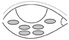
Figure 3
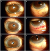
A representative case of conjunctivochalasis surgery. (A, B) A 81-year-old man. Preoperative appearance. (C, D) One week after surgery. (E, F) One month after surgery.


References
1. Meller D, Tseng SC. Conjunctivochalasis: literature review and possible pathophysiology. Surv Ophthalmol. 1998. 43:225–232.
2. Di Pascuale MA, Espana EM, Kawakita T, Tseng SC. Clinical characteristics of conjunctivochalasis with or without aqueous tear deficiency. Br J Ophthalmol. 2004. 88:388–392.
3. Jordan DR, Pelletier CR. Conjunctivochalasis. Can J Ophthalmol. 1996. 31:192–193.
4. Huges WL. Conjunctivochalasis. Am J Ophthalmol. 1942. 25:48–51.
5. Meller D, Maskin SL, Pires RT, Tseng SC. Amniotic membrane transplantation for symptomatic conjunctivochalasis refractory to medical treatments. Cornea. 2000. 19:796–803.
6. Georgiadis NS, Terzidou CD. Epiphora caused by conjunctivochalasis: treatment with transplantation of preserved human amniotic membrane. Cornea. 2001. 20:619–621.
7. Kheirkhah A, Casas V, Blanco G, et al. Amniotic membrane transplantation with fibrin glue for conjunctivochalasis. Am J Ophthalmol. 2007. 144:311–313.
8. Lim HJ, Lee JK, Park DJ. Conjunctivochalasis surgery: amniotic membrane transplantation with fibrin glue. J Korean Ophthalmol Soc. 2008. 49:195–204.
9. Nam K, Jo YJ, Lee SB. The efficacy of fibrin glue in surgical treatment of conjunctivochalasis with epiphora. J Korean Ophthalmol Soc. 2010. 51:498–503.
10. Oh SJ, Byon DS. Treatment of conjunctivochalasis using bipolar cautery. J Korean Ophthalmol Soc. 1999. 40:707–711.
11. Otaka I, Kyu N. A new surgical technique for management of conjunctivochalasis. Am J Ophthalmol. 2000. 129:385–387.
12. Youm DJ, Kim JM, Choi CY. Simple surgical approach with high-frequency radio-wave electrosurgery for conjunctivochalasis. Ophthalmology. 2010. 117:2129–2133.
13. Walt J. Ocular Surface Disease Index (OSDI) Administration and Scoring Manual. 2004. Irvine, CA: Allergan, Inc..
14. Bron AJ, Evans VE, Smith JA. Grading of corneal and conjunctival staining in the context of other dry eye tests. Cornea. 2003. 22:640–650.
15. Francis IC, Chan DG, Kim P, et al. Case-controlled clinical and histopathological study of conjunctivochalasis. Br J Ophthalmol. 2005. 89:302–305.
16. Watanabe A, Yokoi N, Kinoshita S, et al. Clinicopathologic study of conjunctivochalasis. Cornea. 2004. 23:294–298.
17. Meller D, Li DQ, Tseng SC. Regulation of collagenase, stromelysin, and gelatinase B in human conjunctival and conjunctivochalasis fibroblasts by interleukin-1 beta and tumor necrosis factor-alpha. Invest Ophthalmol Vis Sci. 2000. 41:2922–2929.
18. Ko SM, Kim MK, Kim JC. The role of mast cell in hyperlaxity of conjunctiva. J Korean Ophthalmol Soc. 1997. 38:949–955.
19. Udell IJ, Kenyon KR, Sawa M, Dohlman CH. Treatment of superior limbic keratoconjunctivitis by thermocauterization of the superior bulbar conjunctiva. Ophthalmology. 1986. 93:162–166.
 XML Download