

 PDF
PDF ePub
ePub Citation
 Print
Print


Abstract
Purpose
To evaluate the influence of a silicone tube on tear drainage in patients with a healed rhinostomy site after dacryocystorhinostomy.
Methods
The subjects of the present study included the patients for whom the removal of a silicone tube was performed after dacryocystorhinostomy for acquired nasolacrimal duct obstruction. The silicone tube was removed after the rhinostomy site was completely healed. The tear drainage function was evaluated using the fluorescein dye disappearance test at the following 3 time points: immediately before, immediately after, and 1 month after silicone tube removal. In addition, a Schirmer test was performed and tear break-up time was measured at each time point. To study the correlation between the measured values and subjective tearing symptoms, self-report questionnaires were given to each patient at his/her last visit.
Results
The 3 measured values showed no statistical difference between the 3 time points, immediately before, immediately after, and 1 month after silicone tube removal. When the patients were divided into groups according to their subjective symptomatic changes after silicone tube removal, no group showed statistically significant difference in the 3 measured values before, between, and after silicone tube removal.
Figures and Tables
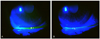 | Figure 1Slit-lamp photography of tear meniscus height using fluorescein dye disappearance test. Tear meniscus height measured immediately after fluorescein dye instillation (A); tear meniscus height measured 5 minutes after fluorescein dye instillation (B); Tear drainage function (%) was defined as (a-b)/a×100 in this study.
|
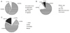 | Figure 2The questionnaire results of the number of patients in each category. (A) The degree of symptomatic improvement after dacryocystorhinostomy. (B) The timing of first symptomatic improvement. (C) Patients' discomfort due to silicone tube.
|
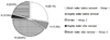 | Figure 3The number of patients according to the symptomatic improvement before and after silicone tube removal. Group 1, patients who felt better symptomatic improvement before silicone tube removal; Group 2, patients who felt similar symptoms before and after silicone tube removal; Group 3, patients who felt better symptomatic improvement after silicone tube removal.
|
Table 1
The changes in tear drainage function and tear film parameters before, immediately after, and 1 month after silicone tube removal

Values are presented as mean ± SD.
TDF = tear drainage function; BUT = tear break-up time.
*Statistical values between before and immediately after silicone tube removal; †Statistical values between immediately after and 1 month after silicone tube removal; ‡Statistical values between before and 1 month after silicone tube removal.
![]()
Table 2
The changes in tear drainage function before and 1 month after silicone tube removal according to the difference in tearing symptoms

![]()
Table 3
The changes in BUT before and 1 month after silicone tube removal according to the difference in tearing symptoms
![]()

References
1. Davies MJ, Lee S, Lemke S, Ghabrial R. Predictors of anatomical patency following primary endonasal dacryocystorhinostomy: a pilot study. Orbit. 2011. 30:49–53.
2. Güler Z, Suat HU, Alp U. Complications and surface reaction associated with silicone intubation. Orbit. 1997. 16:193–199.
3. Smirnov G, Tuomilehto H, Teräsvirta M, et al. Silicone tubing is not necessary after primary endoscopic dacryocystorhinostomy: a prospective randomized study. Am J Rhinol. 2008. 22:214–217.
4. Smirnov G, Tuomilehto H, Teräsvirta M, et al. Silicone tubing after endoscopic dacryocystorhinostomy: is it necessary? Am J Rhinol. 2006. 20:600–602.
5. Charalampidou S, Fulcher T. Does the timing of silicone tube removal following external dacryocystorhinostomy affect patients' symptoms? Orbit. 2009. 28:115–119.
6. Hartikainen J, Grenman R, Puukka P, Seppä H. Prospective randomized comparison of external dacryocystorhinostomy and endonasal laser dacryocystorhinostomy. Ophthalmology. 1998. 105:1106–1113.
7. Onerci M. Dacryocystorhinostomy. Diagnosis and treatment of nasolacrimal canal obstructions. Rhinology. 2002. 40:49–65.
8. Kim CH, Lew H, Yun YS. Correspondence among the canaliculus irrigation test, dacryocystography and Jones test in the epiphora patients. J Korean Ophthalmol Soc. 2007. 48:1017–1022.
9. Joo KS, Lee JK. Comparison of lacrimal scintigraphy and fluorescein dye disappearance test in functional blockage of lacrimal system. J Korean Ophthalmol Soc. 2011. 52:1013–1018.
10. Dolmetsch AM. Nonlaser endoscopic endonasal dacryocystorhinostomy with adjunctive Mitomycin C in nasolacrimal duct obstruction in adults. Ophthalmology. 2010. 117:1037–1040.
11. Woog JJ, Kennedy RH, Custer PL, et al. Endonasal dacryocystorhinostomy: a report by the American Academy of Ophthalmology. Ophthalmology. 2001. 108:2369–2377.
12. Aslan S, Oksuz H, Okuyucu S, et al. Prolene: a novel, cheap, and effective material in dacryocystorhinostomy. Acta Otolaryngol. 2009. 129:755–759.
13. Elmorsy S, Fayek HM. Rubber tube versus Silicone tube at the osteotomy site in external dacryocystorhinostomy. Orbit. 2010. 29:76–82.
14. Mann BS, Wormald PJ. Endoscopic assessment of the dacryocystorhinostomy ostium after endoscopic surgery. Laryngoscope. 2006. 116:1172–1174.
15. Demirci H, Elner VM. Double silicone tube intubation for the management of partial lacrimal system obstruction. Ophthalmology. 2008. 115:383–385.
16. Tucker SM, Linberg JV, Nguyen LL, et al. Measurement of the resistance to fluid flow within the lacrimal outflow system. Ophthalmology. 1995. 102:1639–1645.
17. Kim HG, Im SK, Park HY, Yoon KC. The changes in tear film after dacryocystorhinostomy. J Korean Ophthalmol Soc. 2010. 51:637–641.
 XML Download