

 PDF
PDF ePub
ePub Citation
 Print
Print


Abstract
Purpose
To evaluate the impact of intravitreal bevacizumab injection on visual function and vision-related quality of life (VR-QOL) in patients with branch retinal vein occlusion (BRVO) using the Korean version of the National Eye Institute Visual Function Questionnaire 25 (K-NEI-VFQ-25).
Methods
This study included 32 normal control subjects and 32 patients with BRVO. The Korean version of NEI-VFQ-25 was answered by the patients with BRVO before and 3 months after intravitreal bevacizumab injection, as well as by normal control subjects. Clinical data were collected, including central macular thickness (CMT), total macular volume (TMV) (using time-domain optical coherence tomography [OCT]), and best corrected visual acuity (BCVA).
Results
Visual acuity, CMT, and TMV significantly improved 3 months after intravitreal bevacizumab injections. No bevacizumab-related systemic or ocular adverse effects following intravitreal drug injections were observed. Significant improvement in the VFQ-25 composite score was observed in patients with BRVO. Subscale scores, including general vision, near activities, distance activities, social functioning, mental health, role difficulties, dependency, and peripheral vision, improved after injection. However, subscale scores regarding general health, ocular pain, driving, and color vision did not improve significantly.
Figures and Tables
Table 1
Preoperative characteristics of patients with macular edema due to branch retinal venous occlusion (BRVO) and normal controls
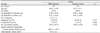
Values are mean ± SD or number unless otherwise indicated.
BCVA = best corrected visual acuity; CMT = central macular thickness (1 mm); log MAR = logarithm of minimum angle of resolution; TMV = total macular volume (6 mm).
*log MAR BCVA of worse-seeing eye of normal controls; †log MAR BCVA of better-seeing eye of normal controls; ‡Data missing for 5 patients and 6 controls; §Data missing for 4 patients and 5 controls.
![]()
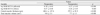
Table 2
Changes in visual acuity and retinal thickness after intravitreal bevacizumab injection in patients with macular edema due to branch retinal venous occlusion (BRVO)
![]()
Table 3
The National Eye Institute 25-item Visual Function Questionnaire (VFQ-25) Composite score and Subscales in Patients with Macular edema due to branch retinal venous occlusion (BRVO) before and after intravitreal bevacizumab injection
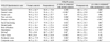
Values are mean ± SD.
VFQ-25 = 25-item Visual Function Questionnaire.
*Significantly different from the normal controls (Mann-Whitney test); †Significantly different from the postoperative values (Wilcoxon signed-rank test); ‡The results of 14 in normal control group and 12 in study group were compared.
![]()
References
1. Rehak J, Rehak M. Branch retinal vein occlusion: pathogenesis, visual prognosis, and treatment modalities. Curr Eye Res. 2008. 33:111–131.
2. Zhao J, Sastry SM, Sperduto RD, et al. The Eye Disease Case-Control Study Group. Arteriovenous crossing patterns in branch retinal vein occlusion. Ophthalmology. 1993. 100:423–428.
3. Lee KS, Chung JK, Lee SJ. Protein C and protein S as a risk factor for retinal vein occlusion. J Korean Ophthalmol Soc. 2005. 46:1796–1801.
4. Silva RM, Faria de Abreu JR, Cunha-Vaz JG. Blood-retina barrier in acute retinal branch vein occlusion. Graefes Arch Clin Exp Ophthalmol. 1995. 233:721–726.
5. The Branch Vein Occlusion Study Group. Argon laser photocoagulation for macular edema in branch vein occlusion. Am J Ophthalmol. 1984. 98:271–282.
6. Lewis H, Schachat AP, Haimann MH, et al. Choroidal neovascularization after laser photocoagulation for diabetic macular edema. Ophthalmology. 1990. 97:503–510. discussion 510-1.
7. Striph GG, Hart WM Jr, Olk RJ. Modified grid laser photocoagulation for diabetic macular edema. The effect on the central visual field. Ophthalmology. 1988. 95:1673–1679.
8. Jonas JB, Akkoyun I, Kamppeter B, et al. Branch retinal vein occlusion treated by intravitreal triamcinolone acetonide. Eye (Lond). 2005. 19:65–71.
9. Lee SJ, Kim ES, Geroski DH, et al. Pharmacokinetics of intraocular drug delivery of Oregon green 488-labeled triamcinolone by subtenon injection using ocular fluorophotometry in rabbit eyes. Invest Ophthalmol Vis Sci. 2008. 49:4506–4514.
10. Rabena MD, Pieramici DJ, Castellarin AA, et al. Intravitreal bevacizumab (Avastin) in the treatment of macular edema secondary to branch retinal vein occlusion. Retina. 2007. 27:419–425.
11. Figueroa MS, Contreras I, Noval S, Arruabarrena C. Results of bevacizumab as the primary treatment for retinal vein occlusions. Br J Ophthalmol. 2010. 94:1052–1056.
12. Jaissle GB, Ziemssen F, Petermeier K, et al. [Bevacizumab for treatment of macular edema secondary to retinal vein occlusion]. Ophthalmologe. 2006. 103:471–475.
13. Shahidi M, Ogura Y, Blair NP, et al. Retinal thickness analysis for quantitative assessment of diabetic macular edema. Arch Ophthalmol. 1991. 109:1115–1119.
14. Park JY, Sung MS, Lee SJ. The focal aggravation of the macular edema on optical coherence tomography after intravitreal triamcinolone injection. J Korean Ophthalmol Soc. 2008. 49:753–762.
15. Mangione CM, Lee PP, Pitts J, et al. Psychometric properties of the National Eye Institute Visual Function Questionnaire (NEI-VFQ). NEI-VFQ Field Test Investigators. Arch Ophthalmol. 1998. 116:1496–1504.
16. Ross CK, Stelmack JA, Stelmack TR, et al. Development and sensitivity to visual impairment of the Low Vision Functional Status Evaluation (LVFSE). Optom Vis Sci. 1999. 76:212–220.
17. Steinberg EP, Tielsch JM, Schein OD, et al. The VF-14. An index of functional impairment in patients with cataract. Arch Ophthalmol. 1994. 112:630–638.
18. Mangione CM, Lee PP, Gutierrez PR, et al. Development of the 25-item National Eye Institute Visual Function Questionnaire. Arch Ophthalmol. 2001. 119:1050–1058.
19. Clemons TE, Chew EY, Bressler SB, McBee W. National Eye Institute Visual Function Questionnaire in the Age-Related Eye Disease Study (AREDS): AREDS Report No. 10. Arch Ophthalmol. 2003. 121:211–217.
20. Klein R, Moss SE, Klein BE, et al. The NEI-VFQ-25 in people with long-term type 1 diabetes mellitus: the Wisconsin Epidemiologic Study of Diabetic Retinopathy. Arch Ophthalmol. 2001. 119:733–740.
21. Rossi GC, Milano G, Tinelli C. The Italian version of the 25-item National Eye Institute Visual Function Questionnaire: translation, validity, and reliability. J Glaucoma. 2003. 12:213–220.
22. Suzukamo Y, Oshika T, Yuzawa M, et al. Psychometric properties of the 25-item National Eye Institute Visual Function Questionnaire (NEI VFQ-25), Japanese version. Health Qual Life Outcomes. 2005. 3:65.
23. Heo JW, Yoon HS, Shin JP, et al. A Validation and Reliability Study of the Korean Version of National Eye Institute Visual Function Questionnaire 25. J Korean Ophthalmol Soc. 2010. 51:1354–1367.
24. Awdeh RM, Elsing SH, Deramo VA, et al. Vision-related quality of life in persons with unilateral branch retinal vein occlusion using the 25-item National Eye Institute Visual Function Questionnaire. Br J Ophthalmol. 2010. 94:319–323.
25. Hamid S, Mirza SA, Shokh I. Branch retinal vein occlusion. J Ayub Med Coll Abbottabad. 2008. 20:128–132.
26. Loftus JV, Sultan MB, Pleil AM. Changes in vision- and health-related quality of life in patients with diabetic macular edema treated with pegaptanib sodium or sham. Invest Ophthalmol Vis Sci. 2011. 52:7498–7505.
27. Ghazi-Nouri SM, Tranos PG, Rubin GS, et al. Visual function and quality of life following vitrectomy and epiretinal membrane peel surgery. Br J Ophthalmol. 2006. 90:559–562.
28. Tranos PG, Ghazi-Nouri SM, Rubin GS, et al. Visual function and subjective perception of visual ability after macular hole surgery. Am J Ophthalmol. 2004. 138:995–1002.
29. Westlake W. Is a one eyed racing driver safe to compete? Formula one (eye) or two? Br J Ophthalmol. 2001. 85:619–624.
30. Cahill MT, Stinnett SS, Banks AD, et al. Quality of life after macular translocation with 360 degrees peripheral retinectomy for age-related macular degeneration. Ophthalmology. 2005. 112:144–151.
31. Curcio CA, Sloan KR, Kalina RE, Hendrickson AE. Human photoreceptor topography. J Comp Neurol. 1990. 292:497–523.
32. Tso MO. Pathology of cystoid macular edema. Ophthalmology. 1982. 89:902–915.
33. Murakami T, Tsujikawa A, Ohta M, et al. Photoreceptor status after resolved macular edema in branch retinal vein occlusion treated with tissue plasminogen activator. Am J Ophthalmol. 2007. 143:171–173.
34. Yamaike N, Tsujikawa A, Sakamoto A, et al. Retinal sensitivity after intravitreal injection of bevacizumab for the treatment of macular edema secondary to retinal vein occlusion. Retina. 2009. 29:757–767.
 XML Download