

 PDF
PDF ePub
ePub Citation
 Print
Print


Abstract
Purpose
To compare changes of anterior and posterior corneal elevation, and sagittal curvature according to the severity of keratoconus and to compare differences between keratoconus and normal cornea.
Methods
A total of 81 eyes diagnosed with keratoconus and 20 eyes of normal subjects were evaluated with a Pentacam Scheimpflug camera. The keratoconus eyes were divided into 3 groups according to mean keratometer (K): mild (K ≤ 47.0 diopters (D)), moderate (47.0 to 52.0 D), and severe (52.0 D≥). The following parameters were obtained to evaluate the correlation of keratoconus: corneal thickness, anterior and posterior corneal elevation, and sagittal curvature.
Results
Out of 81 keratoconus eyes, 56 eyes were mild, 12 eyes were moderate, and 13 eyes were severe keratoconus. The mean central corneal keratometer, anterior and posterior corneal elevation, and sagittal curvature of the keratoconus eyes were 49.7 D, 22.07 µm, 38.16 µm, 52.76 D and the values increased statistically compared to the normal eyes. Furthermore, the values increased significantly with the severity of keratoconus. ROC curve analysis showed the estimated meaningful value for anterior and posterior corneal elevation and sagittal curvature of keratoconus for diagnosis; there were no diagnostic values for corneal thickness and refractive power.
Conclusions
The index of 5.5 µm for mean anterior elevation, 12.5 µm for mean posterior elevation, and 44.5 µm for mean sagittal curvature using the Pentacam® are useful to diagnose keratoconus. Variation of anterior and posterior elevation, and sagittal curvature measured by Pentacam® are useful in understanding the process of keratoconus.
Figures and Tables
Figure 1
Comparison of parameters of Pentacam® (OCULUS, Germany) in keratoconic and normal eyes.(A) Keratometry. (B) Corneal thickness. (C) Anterior and posterior elevation. (D) Sagittal curvature.
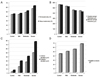
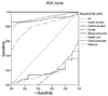
Figure 2
Receiver operating characteristic (ROC) graph showing the sensitivity and specificity of the various corneal measurements. The closer the line to the left-hand side of the graph, the higher the sensitivity and specificity for keratoconus.
References
1. Swartz T, Marten L, Wang M. Measuring the cornea: the latest developments in corneal topography. Curr Opin Ophthalmol. 2007. 18:325–333.
2. Nesburn AB, Bahri S, Salz J, et al. Keratoconus detected by videokeratography in candidates for photorefractive keratectomy. J Refract Surg. 1995. 11:194–201.
3. Fam HB, Lim KL. Corneal elevation indices in normal and keratoconic eyes. J Cataract Refract Surg. 2006. 32:1281–1287.
4. Giers U, Epple C. Comparison of A-scan device accuracy. J Cataract Refract Surg. 1990. 16:235–242.
5. Sonmez B, Doan MP, Hamilton DR. Identification of scanning slit-beam topographic prarameters important in distinguishing normal from keratoconic corneal morphologic features. Am J Ophthalmol. 2007. 143:401–408.
6. de Sanctis U, Missolungi A, Mutani B, et al. Reproducibility and repeatability of central corneal thickness measurement in keratoconus using the rotating Scheimpflug camera and ultrasound pachymetry. Am J Ophthalmol. 2007. 144:712–718.
7. Chen D, Lam AK. Intrasession and intersession repeatability of the Pentacam system on posterior corneal assessment in the normal human eye. J Cataract Refract Surg. 2007. 33:448–454.
8. Lee YJ, Kim SW. The evaluation of enhanced ectasia display mode in screening for keratoconus. J Korean Ophthalmol Soc. 2010. 51:651–657.
9. Lee SU, Lee CH, Lee JE, Lee JS. Corneal topographic study using Orbscan II between keratoconus and keratoconus suspect. J Korean Ophthalmol Soc. 2007. 48:1599–1606.
10. Choi HJ, Kim MK, Lee JL. Diagnostic criteria for keratoconus using Orbscan II slit scanning topography/pachymetry system. J Korean Ophthalmol Soc. 2004. 45:928–935.
11. Rabinowitz YS, McDonnell PJ. Computer-assisted corneal topography in keratoconus. Refract Corneal Surg. 1989. 5:400–408.
12. Roberts C. Characterization of the inherent error in a spherically-biased corneal topography system in mapping a radially aspheric surface. J Refract Corneal Surg. 1994. 10:103–111.
13. Ho JD, Tsai CY, Tsai RJ, et al. Validity of the keratometric index: evaluation by the Pentacam rotating Scheimpflug camera. J Cataract Refract Surg. 2008. 34:137–145.
14. Miháltz K, Kovács I, Takács A, Nagy ZZ. Evaluation of keratometric, pachymetric, and elevation parameters of keratoconic corneas with pentacam. Cornea. 2009. 28:976–980.
15. de Sanctis U, Loiacono C, Richiardi L, et al. Sensitivity and specificity of posterior corneal elevation measured by Pentacam in discriminating keratoconus/subclinical keratoconus. Ophthalmology. 2008. 115:1534–1539.
 XML Download