

 PDF
PDF ePub
ePub Citation
 Print
Print


Abstract
Purpose
To compare central corneal thickness (CCT) as measured by dual rotating Scheimpflug camera (Galilei), anterior segment optical coherence tomography (AS-OCT), and ultrasound pachymetry (USP).
Methods
The measurements of CCT using a dual rotating Scheimpflug camera, AS-OCT, and USP in 40 eyes of 20 healthy subjects were compared.
Results
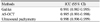
The average measurements of CCT by dual rotating Scheimpflug camera, AS-OCT, and USP were 538.10 ± 31.36 µm, 536.20 ± 31.21 µm, and 541.93 ± 34.93 µm, respectively. The CCT measurement by USP was statistically significantly thicker than by the dual rotating Scheimpflug camera and AS-OCT (p = 0.017, p = 0.001, respectively). There was no significant difference between the dual rotating Scheimpflug camera and AS-OCT (p = 0.054). A significant linear correlation was observed between the dual rotating Scheimpflug camera, the AS-OCT, and the USP (r > 0.900, p < 0.001).
Figures and Tables
Figure 1
Scattergram showing the correlation of central corneal thickness measured by Galilei, anterior segment optical coherence tomography (AS-OCT), and ultrasound pachtmetry (USP). (A) Correlation between Galilei and USP. (B) Correlation between Galilei and AS-OCT. (C) Correlation between AS-OCT and USP.
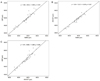
Figure 2
Bland Altman plots between the 2 methods. The middle line is the mean and the lines on the side represent the upper and lower 95% limits of agreement (LoA). (A) Galilei and ultrasound pachymetry (USP). (B) Galilei and anterior segment optical coherence tomography (AS-OCT). (C) AS-OCT and USP.
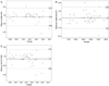
Table 1
Central corneal thickness measured with Galilei, anterior segment optical coherence tomography, and ultrasound pachymetry

References
1. Yau CW, Cheng HC. Microkeratome blades and corneal flap thickness in LASIK. Ophthalmic Surg Lasers Imaging. 2008. 39:471–475.
2. Whitacre MM, Stein RA, Hassanein K. The effect of corneal thickness on applanation tonometry. Am J Ophthalmol. 1993. 115:592–596.
3. Kim DH, Kim MS, Kim JH. Early corneal-thickness changes after penetrating keratoplasty. J Korean Ophthalmol Soc. 1997. 38:1355–1361.
4. Wang Z, Chen J, Yang B. Posterior corneal surface topographic changes after laser in situ keratomileusis are related to residual corneal bed thickness. Ophthalmology. 1999. 106:406–409.
5. Doughty MJ, Zaman ML. Human corneal thickness and its impact on intraocular pressure measures: a review and meta-analysis approach. Surv Ophthalmol. 2000. 44:367–408.
6. Realini T, Lovelace K. Measuring central corneal thickness with ultrasound pachymetry. Optom Vis Sci. 2003. 80:437–439.
7. Sanchis-Gimeno JA, Lleo-Perez A, Casanova J, et al. Inter-observer variability of central corneal thickness measurements using non-contact specular microscopy after laser in situ keratomileusis. Clin Exp Optom. 2004. 87:15–18.
8. Yoo C, Eom YS, Suh YW, Kim YY. Central corneal thickness and anterior scleral thickness in Korean patients with open-angle glaucoma: an anterior segment optical coherence tomography study. J Glaucoma. 2011. 20:95–99.
9. Hashemi H, Roshani M, Mehravaran S, et al. Effect of corneal thickness on the agreement between ultrasound and Orbscan II pachymetry. J Cataract Refract Surg. 2007. 33:1694–1700.
10. Ceylan OM, Turk A, Erdurman C, et al. Comparison of Oculus Pentacam and Stratus optical coherence tomography for measurement of central corneal thickness. Cornea. 2011. 30:670–674.
11. Li EY, Mohamed S, Leung CK, et al. Agreement among 3 methods to measure corneal thickness: ultrasound pachymetry, Orbscan II, and Visante anterior segment optical coherence tomography. Ophthalmology. 2007. 114:1842–1847.
12. Wang L, Shirayama M, Koch DD. Repeatability of corneal power and wavefront aberration measurements with a dual-Scheimpflug Placido corneal topographer. J Cataract Refract Surg. 2010. 36:425–430.
13. Zhao MH, Zou J, Wang WQ, Li J. Comparison of central corneal thickness as measured by non-contact specular microscopy and ultrasound pachymetry before and post LASIK. Clin Experiment Ophthalmol. 2007. 35:818–823.
14. Doughty MJ, Jonuscheit S. An assessment of regional differences in corneal thickness in normal human eyes, using the Orbscan II or ultrasound pachymetry. Optometry. 2007. 78:181–190.
15. Al-Mezaine HS, Al-Amro SA, Kangave D, et al. Comparison of central corneal thickness measurements using Pentacam and ultrasonic pachymetry in post-LASIK eyes for myopia. Eur J Ophthalmol. 2010. 20:852–857.
16. Kim HY, Budenz DL, Lee PS, et al. Comparison of central corneal thickness using anterior segment optical coherence tomography vs ultrasound pachymetry. Am J Ophthalmol. 2008. 145:228–232.
17. Thomas J, Wang J, Rollins AM, Sturm J. Comparison of corneal thickness measured with optical coherence tomography, ultrasonic pachymetry, and a scanning slit method. J Refract Surg. 2006. 22:671–678.
18. Ling T, Ho A, Holden BA. Method of evaluating ultrasonic pachometers. Am J Optom Physiol Opt. 1986. 63:462–466.
19. Copt RP, Thomas R, Mermoud A. Corneal thickness in ocular hypertension, primary open-angle glaucoma, and normal tension glaucoma. Arch Ophthalmol. 1999. 117:14–16.
20. Harper CL, Boulton ME, Bennett D, et al. Diurnal variations in human corneal thickness. Br J Ophthalmol. 1996. 80:1068–1072.
21. Rodrigues EB, Johanson M, Penha FM. Anterior segment tomography with the cirrus optical coherence tomography. J Ophthalmol. 2012. 2012:806989. Epub 2012 Jan 24.
22. Savini G, Carbonelli M, Barboni P, Hoffer KJ. Repeatability of automatic measurements performed by a dual Scheimpflug analyzer in unoperated and post-refractive surgery eyes. J Cataract Refract Surg. 2011. 37:302–309.
23. Yeter V, Sönmez B, Beden U. Comparison of central corneal thickness measurements by Galilei Dual-Scheimpflug analyzer® and ultrasound pachymeter in myopic eyes. Ophthalmic Surg Lasers Imaging. 2012. 43:128–134.
24. Ladi JS, Shah NA. Comparison of central corneal thickness measurements with the Galilei dual Scheimpflug analyzer and ultrasound pachymetry. Indian J Ophthalmol. 2010. 58:385–388.
25. Bechmann M, Thiel MJ, Neubauer AS, et al. Central corneal thickness measurement with a retinal optical coherence tomography device versus standard ultrasonic pachymetry. Cornea. 2001. 20:50–54.
26. Leung DY, Lam DK, Yeung BY, Lam DS. Comparison between central corneal thickness measurements by ultrasound pachymetry and optical coherence tomography. Clin Experiment Ophthalmol. 2006. 34:751–754.
27. Azen SP, Burg KA, Smith RE, Maguen E. A comparison of three methods for the measurement of corneal thickness. Invest Ophthalmol Vis Sci. 1979. 18:535–538.
28. Lee YE, Jun RM. The intra and inter-examiner repeatability of corneal parameters obtained by GALILEI(TM) in normal subjects. J Korean Ophthalmol Soc. 2009. 50:1611–1616.
29. Menassa N, Kaufmann C, Goggin M, et al. Comparison and reproducibility of corneal thickness and curvature readings obtained by the Galilei and the Orbscan II analysis systems. J Cataract Refract Surg. 2008. 34:1742–1747.
 XML Download