

 PDF
PDF ePub
ePub Citation
 Print
Print


Abstract
Purpose
To report a case of unilateral Purtscher's retinopathy that spontaneously resolved within 24 hours.
Case summary
A 54-year-old man presented with decreased visual acuity in his left eye after a vehicle accident. When the accident occurred, his chest region was compressed by the safety belt. The case was diagnosed with Purtscher's retinopathy based on fundus examination, flourescein angiography (FAG) and optical coherent tomography (OCT). At presentation, the best corrected visual acuity (BCVA) was 0.3 in the affected eye. Tiny Purtscher-flecken and macular edema were observed but there was no sign of retinal hemorrhage. Immediately after the trauma, OCT detected abnormally increased hyperreflectivity in the nerve fiber layer and ganglion cell layer, severe cystoid edema and serous foveal detachment. Without any treatment, BCVA was improved to 1.0 within 12 hours. Recovery of visual acuity was followed by improvement of abnormal hyperreflectivity in the nerve fiber layer, cystoid macular edema and serous foveal detachment.
Figures and Tables
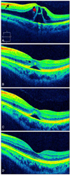
Figure 1
(A) At presentation, severe cystoid macular edema (asterisk) in fovea and hyper-reflectivity (arrow) in the nerve fiber layer and ganglion cell layer. (B) 7 hours later, cystoid edema much improved. (C) 12 hours later, cystoid edema disappeared and only foveal serous detach is shown. (D) 20 hours later, nearly normal OCT finding but mild edema persists in the outer nuclear layer.
References
1. Purtscher O. Noch unbekannte Befunde nach Schädeltrauma. Ber Dtsch Ophthalmol Ges. 1910. 36:294–301.
2. Roncone DP. Purtscher's retinopathy. Optometry. 2002. 73:166–172.
3. Blodi BA, Johnson MW, Gass JD, et al. Purtscher's-like retinopathy after childbirth. Ophthalmology. 1990. 97:1654–1659.
4. Beckingsale AB, Rosenthal AR. Early fundus fluorescein angiographic findings and sequelae in traumatic retinopathy: case report. Br J Ophthalmol. 1983. 67:119–123.
5. Burton TC. Unilateral Purtscher's retinopathy. Ophthalmology. 1980. 87:1096–1105.
6. Fischbein F, Safir A. Monocular Purtscher's retinopathy. A fluorescein angiographic study. Arch Ophthalmol. 1971. 85:480–484.
7. Gomez-Ulla F, Fente B, Torreiro MG, et al. Choroidal vascular abnormality in Purtscher's retinopathy shown by indocyanine green angiography. Am J Ophthalmol. 1996. 122:261–263.
8. Wang AG, Yen MY, Liu JH. Pathogenesis and neuroprotective treatment in Purtscher's retinopathy. Jpn J Ophthalmol. 1998. 42:318–322.
9. Bae JG, Park JS. Purtscher's retinopathy due to safety belts. J Korean Ophthalmol Soc. 2003. 44:231–234.
10. Park JY, Nam WH, Kim SH, et al. Evaluation of the central macula in commotio retinae not associated with other types of traumatic retinopathy. Korean J Ophthalmol. 2011. 25:262–267.
11. Kim SJ, Kim JY, Park SP. Spontaneous resolution of post-traumatic bilateral serous retinal detachment in childrens. J Korean Ophthalmol Soc. 2008. 49:1018–1021.
12. Kaur C, Foulds WS, Ling EA. Blood-retinal barrier in hypoxic ischaemic conditions: basic concepts, clinical features and management. Prog Retin Eye Res. 2008. 27:622–647.
13. Heo J, Lee YC, Yang SW, et al. A case of Purtscher's retinopathy responsive to high-dose steroid therapy. J Korean Ophthalmol Soc. 2008. 49:509–513.
14. Kiryu J, Ogura Y. Hyperbaric oxygen treatment for macular edema in retinal vein occlusion: relation to severity of retinal leakage. Ophthalmologica. 1996. 210:168–170.
 XML Download