

 PDF
PDF ePub
ePub Citation
 Print
Print


Abstract
Purpose
To report a case of hemorrhagic lymphangiectasia treated with surgical excision and confirmed by pathologic examination.
Case summary
A 21-year-old man presented with spontaneous hyperemia of his right eye of 1 week duration. The patient had a history of tuberculous retinal vasculitis and uveitis 1 year prior, but there was no active lesion during regular follow-up. There was no history of trauma, visual disturbance, diplopia, ocular pain, or any sign of systemic disease. Slit lamp examination showed tortuous dilatation of blood-filled lymphatic vessels on temporal conjunctiva of the right eye. The lesion did not change during the 4 weeks of follow-up and local excision biopsy was made for final diagnosis and treatment. Pathologic examinations revealed thin-walled lymphatic vessels with localized dilatation which contained blood in the lumen consistent with hemorrhagic lymphangiectasia. There was no sign of recurrence until 2 months after the operation.
Figures and Tables
Figure 1
Slit lamp photograph shows tortuous dilatation of conjunctival lymphatic vessels which are segmentally filled with blood, with accompanying subconjunctival hemorrhage.
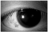
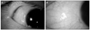
Figure 2
Postoperative slit lamp photographs. (A) Conjunctiva with dilated hemorrhagic lymphatics was excised and the defect was covered with 6.0 × 6.0 mm sized conjunctival autograft. (B) Photograph two months after treatment shows little conjunctival scar but no lymphatic dilatation or subconjunctival hemorrhage.
References
1. Leber T. Lymphangiectasia haemorrhagica conjunctivae. Graefes Arch Ophthalmol. 1880. 26:197–201.
2. Chelsky MP, Magnus DE. Conjunctival hemorrhagic lymphangiectasis. J Am Optom Assoc. 1988. 59:676–678.
3. Scott KR, Tse DT, Kronish JW. Hemorrhagic lymphangiectasia of the conjunctiva. Arch Ophthalmol. 1991. 109:286–287.
4. Jampol LM, Nagpal KC. Hemorrhagic lymphangiectasia of the conjunctiva. Am J Ophthalmol. 1978. 85:419–420.
5. Duke-Elder S, Leigh AG. System of Ophthalmology. Vol. 8. Diseases of the Outer Eye. 1965. St. Louis: CV Mosby;40–42. 1198–1207.
6. Lee SW, Lee SE, Jin KH. Conjunctival inclusion cysts in long-standing chronic vernal keratoconjunctivitis. Korean J Ophthalmol. 2007. 21:251–254.
7. Singh G, Rajaraman R, Raghavan A, Palanisamy M. Bilateral conjunctival retention cysts in the aftermath of Stevens-Johnson syndrome. Indian J Ophthalmol. 2008. 56:70–72.
8. Krachmer JH, Mannis MJ, Holland EJ. Cornea. 2011. Vol. 1:3rd ed. St. Louis: Mosby;488–489.
9. Soong HK, Pollock DA. Hereditary hemorrhagic telangiectasia diagnosed by the ophthalmologist. Cornea. 2000. 19:849–850.
10. Wasfy IA. Lymphangiectasia haemorrhagica conjunctivae: report of three cases with a note on successful treatment with cryosurgery in one case. Bull Ophthalmol Soc Egypt. 1975. 68:37–44.
 XML Download