

 PDF
PDF ePub
ePub Citation
 Print
Print


Abstract
Purpose
To compare the visual field and retinal nerve fiber layer of scleral buckling (SB) and primary pars plana vitrectomy (PPV) for treatment of simple rhegmatogenous retinal detachment (RRD).
Methods
We studied 20 eyes with RRD that were underwent successful surgical reattachment. Visual field test and retinal nerve fiber layer (RNFL) thickness measurements were performed in patients, and outcomes were compared not only between the operated eye and fellow eye, but also between SB and PPV 3 months postoperatively.
Results
After the operation, PSD and MD were higher in the operated eye than in the fellow eye (p = 0.002, p < 0.001, respectively). RNFL thickness was lower in the operated eye than in the fellow eye (p < 0.001). No significant differences in BCVA were detected between SB and PPV. However, the respective differences between the operated eye and fellow eye regarding pattern standard deviation (4.0 ± 4.0, 0.7 ± 1.5), mean deviation (6.5 ± 4.6, 1.9 ± 1.9), and RNFL (8.2 ± 10.3 µm, 1.8 ± 2.7 µm) were significantly higher in PPV than in SB.
Figures and Tables
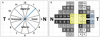 | Figure 1(A) Distribution of superior, nasal, inferior, temporal sectors in RNFL scan. (B) Distribution of VF zones and associated HVF central 24-2 test points. Superior RNFL thickness associated 3, 4, 5, 6, 7, 8 VF test points. Temporal RNFL thickness associated 9, 10, 12, 13 VF test points (yellow box). Inferior RNFL thickness associated 14, 15, 16, 17, 18, 19, 20 VF test points. Nasal RNFL thickness associated 1, 21 VF test points (blue box). N = nasal; T = temporal.
|
Table 3
Comparison of vitrectomized eyes and scleral buckling eyes 3 months after operation
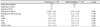
Values are presented as mean ± SD or number.
PCO = posterior capsule opacity; ΔPSD = difference between PSD of the eye with operation and the fellow eye; ΔMD = difference between MD of the eye with operation and fellow eye; ΔRNFL = difference between retinal nerve fiber layer of the eye with operation and the fellow eye.
*Mann-Whitney U-test.
![]()
References
1. Ah-Fat FG, Sharma MC, Majid MA, et al. Trends in vitreoretinal surgery at a tertiary referral centre: 1987 to 1996. Br J Ophthalmol. 1999. 83:396–398.
2. Koh TH, Choi MJ, Cho SW, et al. Scleral buckling and primary vitrectomy in simple rhegmatogenous retinal detachment. J Korean Ophthalmol Soc. 2010. 51:366–371.
3. Yan H, Dhurjon L, Chow DR, et al. Visual field defect after pars plana vitrectomy. Ophthalmology. 1998. 105:1612–1616.
4. Sato EA, Shinoda K, Inoue M, et al. Reduced choroidal blood flow can induce visual field defect in open angle glaucoma patients without intraocular pressure elevation following encircling scleral buckling. Retina. 2008. 28:493–497.
5. Sasoh M, Ito Y, Wakitani Y, et al. 10-year follow-up of visual functions in patients who underwent scleral buckling. Retina. 2005. 25:965–971.
6. Arantes TE, Garcia CR, Tavares IM, et al. Relationship between retinal nerve fiber layer and visual field function in human immunodeficiency virus-infected patients without retinitis. Retina. 2012. 32:152–159.
7. Weber J, Ulrich H. A perimetric nerve fiber bundle map. Int Ophthalmol. 1991. 15:193–200.
8. Heimann H, Bartz-Schmidt KU, Bornfeld N, et al. Scleral buckling versus primary vitrectomy in rhegmatogenous retinal detachment: a prospective randomized multicenter clinical study. Ophthalmology. 2007. 114:2142–2154.
9. Garweg JG, Bergstein D, Windisch B, et al. Recovery of visual field and acuity after removal of epiretinal and inner limiting membranes. Br J Ophthalmol. 2008. 92:220–224.
10. Chong DY, Fuller DG. The declining use of scleral buckling with vitrectomy for primary retinal detachments. Arch Ophthalmol. 2010. 128:1206–1207.
11. Kim YK, Woo SJ, Park KH, et al. Comparison of persistent submacular fluid in vitrectomy and scleral buckle surgery for macula-involving retinal detachment. Am J Ophthalmol. 2010. 149:623–629.
12. Fisher SK, Lewis GP. Müller cell and neuronal remodeling in retinal detachment and reattachment and their potential consequences for visual recovery: a review and reconsideration of recent data. Vision Res. 2003. 43:887–897.
13. Kerrison JB, Haller JA, Elman M, Miller NR. Visual field loss following vitreous surgery. Arch Ophthalmol. 1996. 114:564–569.
14. Hirata A, Yonemura N, Hasumura T, et al. Effect of infusion air pressure on visual field defects after macular hole surgery. Am J Ophthalmol. 2000. 130:611–616.
15. Yang SS, McDonald HR, Everett AI, et al. Retinal damage caused by air-fluid exchange during pars plana vitrectomy. Retina. 2006. 26:334–338.
16. Ezra E, Arden GB, Riordan-Eva P, et al. Visual field loss following vitrectomy for stage 2 and 3 macular holes. Br J Ophthalmol. 1996. 80:519–525.
17. Nagahara M, Tamaki Y, Araie M, Eguchi S. Effects of scleral buckling and encircling procedures on human optic nerve head and retinochoroidal circulation. Br J Ophthalmol. 2000. 84:31–36.
18. Ozdek S, Lonneville Y, Onol M, et al. Assessment of retinal nerve fiber layer thickness with NFA-GDx following successful scleral buckling surgery. Eur J Ophthalmol. 2003. 13:697–701.
 XML Download