

 PDF
PDF ePub
ePub Citation
 Print
Print


Abstract
Purpose
To analyze changes of higher-order aberrations (HOAs) after micro-coaxial cataract surgery according to pupil size and to evaluate systemic factors affecting these changes.
Methods
Forty-two patients (42 eyes) who had undergone micro-coaxial cataract surgery were followed-up in the present study. HOAs (total RMS, coma, trefoil and spherical aberration) were measured at 1 week, 1 month, 3 months and 6 months post surgery. Differences of HOAs according to pupil size and systemic factors affecting changes of HOAs were analyzed.
Results
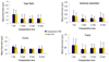
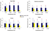
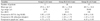
Total RMS (1.08 ± 0.89 µm) and spherical aberration (0.03 ± 0.13 µm) were lower in the group with a pupil size between 5.6 and 6.4 mm at 1 month after surgery compared with the group with pupil size lower than 5.5 mm (1.69 ± 0.97 µm, 0.09 ± 0.11 µm) or the group with pupil size larger than 6.5 mm (1.75 ± 0.87 µm, 0.12 ± 0.18 µm), which remained low at 6 months after surgery. Coma and trefoil showed a similar tendency. Patients with diabetes mellitus had smaller pupil size and higher total RMS, coma, trefoil and spherical aberration values at 1 month after surgery compared with patients without diabetes.
Figures and Tables
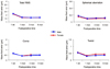
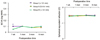 | Figure 1The changes of BCVA (Best corrected visual acuity) and spherical equivalent refraction after microcoaxial cataract surgery in the 3 groups according to pupil size (*p < 0.05).
|
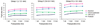 | Figure 2Postoperative pupil size in 3 groups checked by Wavefront®, Colvard pupillometer®, ORB scan II® and Pentacam®.
|
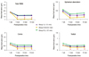 | Figure 3The changes of higher-order aberrations after microcoaxial cataract surgery in the 3 groups according to pupil size
(*p < 0.05, compared with group I or group III).
|
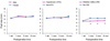 | Figure 4Postoperative pupil size measured by Colvard pupillometer® according to sex, the presence of hypertension and diabetic mellitus (*p < 0.05, compared with non-DM group).
|
 | Figure 5The changes of higher-order aberrations after microcoaxial cataract surgery according to sex.
|
 | Figure 6The changes of higher-order aberrations after microcoaxial cataract surgery according to the presence of hypertension.
|
 | Figure 7The changes of higher-order aberrations after microcoaxial cataract surgery according to the presence of diabetic mellitus (*p < 0.05, compared with non-DM group).
|
References
1. Kelman CD. Phaco-emulsification and aspiration. A new technique of cataract removal. A preliminary report. Am J Ophthalmol. 1967. 64:23–35.
2. Vasavada V, Vasavada V, Raj SM, Vasavada AR. Intraoperative performance and postoperative outcomes of microcoaxial phacoemulsification. Observational study. J Cataract Refract Surg. 2007. 33:1019–1024.
3. Osher RH, Injev VP. Microcoaxial phacoemulsification Part 1: laboratory studies. J Cataract Refract Surg. 2007. 33:401–407.
4. Chalita MR, Krueger RR. Correlation of aberrations with visual acuity and symptoms. Ophthalmol Clin North Am. 2004. 17:135–142.
5. Williams D, Yoon GY, Porter J, et al. Visual benefit of correcting higher order aberrations of the eye. J Refract Surg. 2000. 16:S554–S559.
6. Werner L, Mamalis N. Wavefront corrections of intraocular lenses. Ophthalmol Clin North Am. 2004. 17:233–245.
7. Guirao A, Redondo M, Geraghty E, et al. Corneal optical aberrations and retinal image quality in patients in whom monofocal intraocular lenses were implanted. Arch Ophthalmol. 2002. 120:1143–1151.
8. Kawamorita T, Uozato H, Handa T, et al. Effect of pupil size on visual acuity in a laboratory model of pseudophakic monovision. J Refract Surg. 2010. 26:378–380.
9. Wang Y, Zhao K, Jin Y, et al. Changes of higher order aberration with various pupil sizes in the myopic eye. J Refract Surg. 2003. 19:S270–S274.
10. Mun GH, Im SK, Park HY, Yoon KC. Comparison of visual function between two aspheric intraocular lenses after microcoaxial cataract surgery. J Korean Ophthalmol Soc. 2010. 51:333–339.
11. Kasper T, Bühren J, Kohnen T. Intraindividual comparison of higher-order aberrations after implantation of aspherical and spherical intraocular lenses as a function of pupil diameter. J Cataract Refract Surg. 2006. 32:78–84.
12. Caporossi A, Martone G, Casprini F, Rapisarda L. Prospective randomized study of clinical performance of 3 aspheric and 2 spherical intraocular lenses in 250 eyes. J Refract Surg. 2007. 23:639–648.
13. Packer M, Fine IH, Hoffman RS. Aspheric intraocular lens selection: the evolution of refractive cataract surgery. Curr Opin Ophthalmol. 2008. 19:1–4.
14. Hayashi K, Hayashi H. Stereopsis in bilaterally pseudophakic patients. J Cataract Refract Surg. 2004. 30:1466–1470.
15. Ko BU, Ryu WY, Park WC. Pupil size in the normal korean population according to age and illuminance. J Korean Ophthalmol Soc. 2011. 52:401–406.
16. Kohnen T, Terzi E, Kasper T, et al. Correlation of infrared pupillometers and CCD-camera imaging from aberrometry and videokeratography for determining scotopic pupil size. J Cataract Refract Surg. 2004. 30:2116–2123.
17. Yazici AT, Bozkurt E, Alagoz C, et al. Central corneal thickness, anterior chamber depth, and pupil diameter measurements using Visante OCT, Orbscan, and Pentacam. J Refract Surg. 2010. 26:127–133.
18. Gibbens MV, Goel R, Smith SE. Effect of cataract extraction on the pupil response to mydriatics. Br J Ophthalmol. 1989. 73:563–565.
19. Yap EY, Aung T, Fan RF. Pupil abnormalities on the first postoperative day after cataract surgery. Int Ophthalmol. 1996-1997. 20:187–192.
20. Koch DD, Samuelson SW, Villarreal R, et al. Changes in pupil size induced by phacoemulsification and posterior chamber lens implantation: consequences for multifocal lenses. J Cataract Refract Surg. 1996. 22:579–584.
21. Hayashi K, Hayashi H. Pupil size before and after phacoemulsification in nondiabetic and diabetic patients. J Cataract Refract Surg. 2004. 30:2543–2550.
22. Castejón-Mochón JF, López-Gil N, Benito A, Artal P. Ocular wave-front aberration statistics in a normal young population. Vision Res. 2002. 42:1611–1617.
23. de Castro LE, Sandoval HP, Bartholomew LR, et al. High-order aberrations and preoperative associated factors. Acta Ophthalmol Scand. 2007. 85:106–110.
 XML Download