

 PDF
PDF ePub
ePub Citation
 Print
Print


Abstract
Purpose
To evaluate the long-term changes in the corneal sensitivity and to analyze which tear film parameters correlate with corneal sensitivity after penetrating keratoplasty (PKP).
Methods
Twenty-eight eyes of 28 patients who underwent PKP were included in the present study. Corneal sensitivity, Schirmer test, tear break-up time, tear clearance rate, and keratoepitheliopathy were evaluated at 1, 3, 6, 12, and 24 months postoperatively. Then, which tear film parameters correlated with corneal sensitivity during the follow-up period were analyzed.
Results
Corneal sensitivity was 5.18 ± 3.96 mm at one month postoperatively and increased to 29.64 ± 12.39 mm (p < 0.05) at 24 months postoperatively but was still not in the normal range. Tear break-up time and keratoepitheliopathy improved at postoperative three months and six months, respectively, compared with one month postoperatively, and both measures normalized at 24 months postoperatively. Schirmer test results and tear clearance rate were in the normal range during the follow-up period. Corneal sensitivity was correlated with tear break-up time from three to 24 months postoperative and with tear clearance rate from one month to 24 months postoperative (p < 0.05).
Figures and Tables
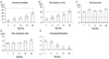
Figure 1
Changes in corneal sensitivity (A), tear film and ocular surface parameters including tear break-up time (B), Schirmer test (C), tear clearance rate (D), and keratoepitheliopathy (E) after penetrating keratoplasty. Dotted line indicates the lowest value of the normal range (*p < 0.05, compared with 1 month data).


References
1. Benitez-del-Castillo JM, del Rio T, Iradier T, et al. Decrease in tear secretion and corneal sensitivity after laser in situ keratomileusis. Cornea. 2001. 20:30–32.
2. Horwath-Winter J, Vidic B, Schwantzer G, Schmut O. Early changes in corneal sensation, ocular surface integrity, and tear-film function after laser-assisted subepithelial keratectomy. J Cataract Refract Surg. 2004. 30:2316–2321.
3. Cho YK, Kim MS. Dry eye after cataract surgery and associated intraoperative risk factors. Korean J Ophthalmol. 2009. 23:65–73.
4. Kim JH, Sah WJ, Hahn TW, Lee YC. Some problems after photorefractive keratectomy. J Refract Corneal Surg. 1994. 10:2 Suppl. S226–S230.
5. Vroman DT, Sandoval HP, Fernández de Castro LE, et al. Effect of hinge location on corneal sensation and dry eye after laser in situ keratomileusis for myopia. J Cataract Refract Surg. 2005. 31:1881–1887.
6. Mian SI, Li AY, Dutta S, et al. Dry eyes and corneal sensation after laser in situ keratomileusis with femtosecond laser flap creation Effect of hinge position, hinge angle, and flap thickness. J Cataract Refract Surg. 2009. 35:2092–2098.
7. Situ P, Simpson TL, Fonn D, Jones LW. Conjunctival and corneal pneumatic sensitivity is associated with signs and symptoms of ocular dryness. Invest Ophthalmol Vis Sci. 2008. 49:2971–2976.
8. Stern ME, Beuerman RW, Fox RI, et al. The pathology of dry eye: the interaction between the ocular surface and lacrimal glands. Cornea. 1998. 17:584–589.
9. Lawrenson JG. Corneal sensitivity in health and disease. Ophthalmic Physiol Opt. 1997. 17:suppl 1. S17–S22.
10. Bao L, Wang C, Yang X. [An experimental study on corneal nerve regeneration after penetrating keratoplasty with long-term cryopreserved rabbit corneas]. Zhonghua Yan Ke Za Zhi. 1996. 32:379–381.
11. Ceccuzzi R, Zanardi A, Fiorentino A, et al. Corneal sensitivity in keratoconus after penetrating and deep anterior lamellar keratoplasty. Ophthalmologica. 2010. 224:247–250.
12. Skriver K. Reinnervation of the corneal graft. Acta Ophthalmol (Copenh). 1978. 56:1013–1015.
13. Richter A, Slowik C, Somodi S, et al. Corneal reinnervation following penetrating keratoplasty--correlation of esthesiometry and confocal microscopy. Ger J Ophthalmol. 1996. 5:513–517.
14. Rao GN, John T, Ishida N, Aquavella JV. Recovery of corneal sensitivity in grafts following penetrating keratoplasty. Ophthalmology. 1985. 92:1408–1411.
15. Dogru M, Katakami C, Inoue M. Tear function and ocular surface changes in noninsulin-dependent diabetes mellitus. Ophthalmology. 2001. 108:586–592.
16. Yoon KC, Im SK, Park YG, et al. Application of umbilical cord serum eyedrops for the treatment of dry eye syndrome. Cornea. 2006. 25:268–272.
17. Yoon KC, Im SK, Seo MS. Changes of tear film and ocular surface in diabetes mellitus. Korean J Ophthalmol. 2004. 18:168–174.
18. Yoon KC, Song BY, Seo MS. Effects of smoking on tear film and ocular surface. Korean J Ophthalmol. 2005. 19:18–22.
19. Yoon KC, Park CS, You IC, et al. Expression of CXCL9, -10, -11, and CXCR3 in the tear film and ocular surface of patients with dry eye syndrome. Invest Ophthalmol Vis Sci. 2010. 51:643–650.
20. Yoon KC, Jeong IY, Park YG, Yang SY. Interleukin-6 and tumor necrosis factor-alpha levels in tears of patients with dry eye syndrome. Cornea. 2007. 26:431–437.
21. Yoon KC, Im SK, Kim HG, You IC. Usefulness of double vital staining with 1% fluorescein and 1% lissamine green in patients with dry eye syndrome. Cornea. 2011. 30:972–976.
22. The definition and classification of dry eye disease: report of The Definition and Classification Subcommittee of the International Dry Eye WorkShop (2007). Ocul Surf. 2007. 5:75–92.
23. de Pinho Tavares F, Fernandes RS, Bernardes TF, et al. Dry eye disease. Semin Ophthalmol. 2010. 25:84–93.
24. Müller LJ, Marfurt CF, Kruse F, Tervo TM. Corneal nerves: structure, contents and function. Exp Eye Res. 2003. 76:521–542.
25. Rózsa AJ, Beuerman RW. Density and organization of free nerve endings in the corneal epithelium of the rabbit. Pain. 1982. 14:105–120.
26. Müller LJ, Pels L, Vrensen GF. Ultrastructural organization of human corneal nerves. Invest Ophthalmol Vis Sci. 1996. 37:476–488.
27. Mathers WD, Jester JV, Lemp MA. Return of human corneal sensitivity after penetrating keratoplasty. Arch Ophthalmol. 1988. 106:210–211.
28. Ruben M, Colebrook E. Keratoplasty sensitivity. Br J Ophthalmol. 1979. 63:265–267.
29. Darwish T, Brahma A, Efron N, O'Donnell C. Subbasal nerve regeneration after penetrating keratoplasty. Cornea. 2007. 26:935–940.
30. Battat L, Macri A, Dursun D, Pfluqfelder SC. Effects of laser in situ keratomileusis on tear production, clearance, and the ocular surface. Ophthalmology. 2001. 108:1230–1235.
31. Lee SJ, Kim JK, Seo KY, et al. Comparison of corneal nerve regeneration and sensitivity between LASIK and laser epithelial keratomileusis (LASEK). Am J Ophthalmol. 2006. 141:1009–1015.
32. Afonso AA, Monroy D, Stern ME, et al. Correlation of tear fluorescein clearance and Schirmer test scores with ocular irritation symptoms. Ophthalmology. 1999. 106:803–810.
33. Macri A, Rolando M, Pflugfelder S. A standardized visual scale for evaluation of tear fluorescein clearance. Ophthalmology. 2000. 107:1338–1343.
34. Shimazaki J, Shimmura S, Mochizuki K, Tsubota K. Morphology and barrier function of the corneal epithelium after penetrating keratoplasty: association with original diseases, tear function, and suture removal. Cornea. 1999. 18:559–564.
35. Gong XM. [Recovery of corneal sensitivity following keratoplasty and corneal diseases]. Zhonghua Yan Ke Za Zhi. 1992. 28:150–152.
 XML Download