

 PDF
PDF ePub
ePub Citation
 Print
Print


Abstract
Purpose
We report a case of a full-thickness macular hole treated in a female adult with bilateral retinal capillary hemangiomas.
Case summary
A 20-year-old woman with bilateral retinal capilliary hemangiomas presented with blurred vision in her right eye. A thin epiretinal membrane and impending macular hole were found that did not appear to be related with a 2-disc-diameter-sized retinal angioma, telangiectactic vessels, and hard exudates in the lower retinal area of her right eye. Four months later, optical coherence tomography revealed a full-thickness macular hole in her right eye. A vitrectomy was performed, and the full-thickness macular hole was completely resolved.
Figures and Tables
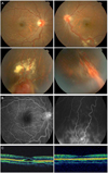
Figure 1
Fundus photographs of both eyes (A) and fluorescein angiographic findings of the right eye (B) at the initial visit revealed retinal angiomas and retinal vascular telangiectactic lesions with exudations in the lower retinal area. Optical coherence tomograph (OCT) of the right eye revealed epiretinal membrane and sensory retinal detachment and OCT of the left eye revealed flat macular finding at the initial visit (C).
References
1. Ridley M, Green J, Johnson G. Retinal angiomatosis: the ocular manifestations of von Hippel-Lindau disease. Can J Ophthalmol. 1986. 21:276–283.
2. Wing GL, Weiter JJ, Kelly PJ, et al. Von Hippel-Lindau disease: angiomatosis of the retina and central nervous system. Ophthalmology. 1981. 88:1311–1314.
3. Raju B, Majji AB, Jalali S. von Hippel angioma in South Indian subjects--a clinical study. Retina. 2003. 23:670–674.
4. Inoue M, Yamazaki K, Shinoda K, et al. A clinicopathologic case report on macular hole associated with von Hippel-Lindau disease: a novel ultrastructural finding of wormlike, wavy tangles of filaments. Graefes Arch Clin Exp Ophthalmol. 2004. 242:881–886.
5. Carr RE, Noble KG. Retinal angiomatosis. Ophthalmology. 1980. 87:956–959. 961
6. Singh A, Shields J, Shields C. Solitary retinal capillary hemangioma: hereditary (von Hippel-Lindau disease) or nonhereditary? Arch Ophthalmol. 2001. 119:232–234.
7. Schwartz PL, Fastenberg DM, Shakin JL. Management of macular puckers associated with retinal angiomas. Ophthalmic Surg. 1990. 21:550–556.
8. Takahashi A, Nagaoka T, Ishiko S, et al. Foveal anatomic changes in a progressing stage 1 macular hole documented by spectral-domain optical coherence tomography. Ophthalmology. 2010. 117:806–810.
9. Schwartz PL, Trubowitsch G, Fastenberg DM, Stein M. Macular pucker and retinal angioma. Ophthalmic Surg. 1987. 18:677–679.
10. McDonald HR, Schatz H, Johnson RN, et al. Vitrectomy in eyes with peripheral retinal angioma associated with traction macular detachment. Ophthalmology. 1996. 103:329–335.
 XML Download