

 PDF
PDF ePub
ePub Citation
 Print
Print


Abstract
Purpose
To compare the fluorescein angiographic findings of nonarteritic anterior ischemic optic neuropathy (NA-AION) and optic neuritis.
Methods
The present study included 41 patients (41 eyes), who were diagnosed with NA-AION or optic neuritis and underwent fluorescein angiography in our clinic. The clinical profiles of patients, characteristics of optic disc head and hemorrhage, and visual field findings were analyzed retrospectively. The onset and filling time, perfusion time of retinal artery, optic disc, and peripapillary choroid were evaluated quantitatively.
Results
Patients with NA-AION showed statistically significant delay in both the onset time, filling time and perfusion time of the optic disc and peripapillary choroid compared with patients with optic neuritis (p < 0.05). There was no significant difference in the dye leakage of the peripapillary areas between the 2 groups (p = 0.324).
Conclusions
In the present study, the results of fluorescein filling were significantly different between the NA-AION group and the optic neuritis group. The results may help determine the therapeutic plan and to differentiate between the 2 disease entities, especially in cases of overlapping clinical features.
Figures and Tables
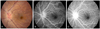 | Figure 1Fluorescein angiographic findings of typical case of optic neuritis. Fundus color photograph (A) shows optic disc edema. The fluorescein angiographic finding at 13 seconds (B) shows the beginning of the filling of optic disc head and peripapillary choroid. The finding at 17 seconds (C) shows the complete filling of peripapillary choroid and optic disc head.
|
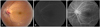 | Figure 2Fluorescein angiographic findings of typical case of non-arteritic anterior ischemic optic neuropathy. Fundus color photograph (A) shows optic disc edema with hemorrhage. The fluorescein angiographic finding at 20 seconds (B) shows the delayed filling of peripapillary choroid and optic disc head. The finding at 36 seconds (C) shows complete filling of peripapillary choroid and optic disc.
|
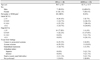
Table 1
Summary of sex, age, periocular pain, initial & final BCVA, and visual field findings in the two group
![]()

References
1. Hayreh SS, Joos KM, Podhajsky PA, Long CR. Systemic diseases associated with nonarteritic anterior ischemic optic neuropathy. Am J Ophthalmol. 1994. 118:766–780.
2. Ellenberger C Jr. Ischemic optic neuropathy as a possible early complication of vascular hypertension. Am J Ophthalmol. 1979. 88:1045–1051.
3. Tomsak RL, Remler BF. Anterior ischemic optic neuropathy and increased intraocular pressure. J Clin Neuroophthalmol. 1989. 9:116–118.
4. Beck RW, Savino PJ, Repka MX, et al. Optic disc structure in anterior ischemic optic neuropathy. Ophthalmology. 1984. 91:1334–1337.
5. Doro S, Lessell S. Cup-disc ratio and ischemic optic neuropathy. Arch Ophthalmol. 1985. 103:1143–1144.
6. Guyer DR, Miller NR, Auer CL, Fine SL. The risk of cerebrovascular and cardiovascular disease in patients with anterior ischemic optic neuropathy. Arch Ophthalmol. 1985. 103:1136–1142.
7. Ahn BC, Kim HS, Ahn HS. Clinical profile of the Optic neuritis in Korea. J Korean Ophthalmol Soc. 1997. 38:1827–1833.
8. Optic Neuritis Study Group. The clinical profile of optic neuritis. Experience of the Optic Neuritis Treatment Trial. Arch Ophthalmol. 1991. 109:1673–1678.
9. Rizzo JF 3rd, Lessell S. Risk of developing multiple sclerosis after uncomplicated optic neuritis: a long-term prospective study. Neurology. 1988. 38:185–190.
10. Park WC, Chang BL. Clinical features of anterior ischemic optic neuropathy. J Korean Ophthalmol Soc. 2003. 44:144–149.
11. Rizzo JF 3rd, Lessell S. Optic neuritis and ischemic optic neuropathy. Overlapping clinical profiles. Arch Ophthalmol. 1991. 109:1668–1672.
12. Arnold AC, Hepler RS. Natural history of nonarteritic anterior ischemic optic neuropathy. J Neuroophthalmol. 1994. 14:66–69.
13. Beck RW, Cleary PA. Optic neuritis treatment trial. One-year follow-up results. Arch Ophthalmol. 1993. 111:773–775.
14. Boghen DR, Glaser JS. Ischaemic optic neuropathy. The clinical profile and history. Brain. 1975. 98:689–708.
15. Jung JY, Kim JS. Comparison of optic disc appearance in anterior ischemic optic neuropathy and optic neuritis. J Korean Ophthalmol Soc. 2003. 44:157–161.
16. Ischemic Optic Neuropathy Decompression Trial Research Group. Characteristics of patients with nonarteritic anterior ischemic optic neuropathy eligible for the Ischemic Optic Neuropathy Decompression Trial. Arch Ophthalmol. 1996. 114:1366–1374.
17. Kim DH, Hwang JM. Risk factors for Korean patients with anterior ischemic optic neuropathy. J Korean Ophthalmol Soc. 2007. 48:1527–1531.
18. Hayreh SS. Anterior ischaemic optic neuropathy. II. Fundus on ophthalmoscopy and fluorescein angiography. Br J Ophthalmol. 1974. 58:964–980.
19. Hayreh SS. Anterior ischaemic optic neuropathy. Differentiation of arteritic from non-arteritic type and its management. Eye. 1990. 4(Pt 1):25–41.
20. Arnold AC, Hepler RS. Fluorescein angiography in acute nonarteritic anterior ischemic optic neuropathy. Am J Ophthalmol. 1994. 117:222–230.
21. Anmarkrud N. The value of fluorescein fundus angiography in evaluating optic disc oedema. Acta Ophthalmol (Copenh). 1977. 55:605–615.
22. Mack HG, O'Day J, Currie JN. Delayed choroidal perfusion in giant cell arteritis. J Clin Neuroophthalmol. 1991. 11:221–227.
23. Siatkowski RM, Gass JD, Glaser JS, et al. Fluorescein angiography in the diagnosis of giant cell arteritis. Am J Ophthalmol. 1993. 115:57–63.
24. Evans PY, Shimizu K, Limaye S, et al. Fluorescein cineangiography of the optic nerve head. Trans Am Acad Ophthalmol Otolaryngol. 1973. 77:OP260–OP273.
25. Schatz H. Ryan SJ, editor. Fluorescein angiography: basic principles and interpretation. Retina. 2006. v. 2:4th ed. St. Louis: Mosby;873–916. chap. 51.
 XML Download