

 PDF
PDF ePub
ePub Citation
 Print
Print


Abstract
Purpose
To evaluate the crystalline lens thickness/anterior chamber depth (CLT/ACD) ratio as a preoperative factor that
affects the vault after implantable contact lens (ICL) implantation.
Methods
A total of 130 eyes of 130 patients who received bilateral ICL implantation were included in the present study.
The preoperative CLT/ACD ratio was analyzed to determine if the patients had any correlation with postoperative vault by
Visante optical coherence tomography (OCT).
Results
The mean vault was 0.58 mm ± 0.23 at postoperative 2 months. Eight eyes (6.15%) had low vault, 93 eyes (71.53%) had ideal vault and 29 eyes (29.31%) had high vault. The CLT/ACD ratios were 1.04 mm ± 0.11, 0.96 mm ± 0.09 and 0.90 mm ± 0.09 in the low vault group, ideal vault group and high vault group, respectively (p < 0.01). The CLT/ACD ratio showed statistically significant correlations with postoperative vault in univariate analysis (r = -0.4718; p < 0.01) and in multivariate analysis (p < 0.01).
Figures and Tables
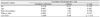
Figure 1
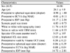
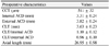
An example of preoperative ultrasound A scan (US-1800, Nidek Inc., Japan) result showing axial parameters including ACD (anterior chamber depth) and Lens (crystalline lens thickness) of both eyes. In this example, the lens thickness to external ACD of the right eye is 1.04 (3.65 mm/3.51 mm).
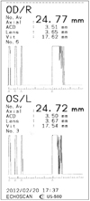
Figure 2
Scatter diagrams with regression line showing the negative correlation of preoperative lens thickness/external anterior chamber depth and vault by Visante OCT. POD = postoperative day; ACD = anterior chamber depth; OCT = optical coherence tomography.
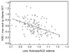
Figure 3
Box-and-whisker plot (A) and dots plot (B) showing the distribution of preoperative lens thickness/external anterior chamber depth according to postoperative vault, by which 3 groups were made. That is low vault group (<250 µm), ideal vault group (250 µm-750 µm) and high vault group (>750 µm). ACD = anterior chamber depth.
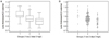
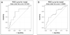
Figure 4
ROC (receiver operating characteristic) curve of preoperative crystalline lens thickness/anterior chamber depth between low vault versus ideal vault group (A), and between high vault versus ideal vault (B).

References
1. Han SY, Lee KH. Long term effect of ICL implantation to treat high myopia. J Korean Ophthalmol Soc. 2007. 48:465–472.
2. Han SY, Moon SJ, Kim HS, et al. Intraindividual comparison of ICL and toric ICL implantation in the correction of high myopia with astigmatism. J Korean Ophthalmol Soc. 2010. 51:802–808.
3. DU GP, Huang YF, Wang LQ, et al. [Outcome after treatment of myopia with implantable Collamer lens]. Zhonghua Yan Ke Za Zhi. 2011. 47:146–150.
4. Pitault G, Leboeuf C, Leroux Les Jardins S, et al. [Ultrasound biomicroscopy of posterior chamber phakic intraocular lenses: a comparative study between ICL and PRL models]. J Fr Ophtalmol. 2005. 28:914–923.
5. Alfonso JF, Lisa C, Abdelhamid A, et al. Posterior chamber phakic intraocular lenses after penetrating keratoplasty. J Cataract Refract Surg. 2009. 35:1166–1173.
6. Fernandes P, González-Méijome JM, Madrid-Costa D, et al. Implantable collamer posterior chamber intraocular lenses: a review of potential complications. J Refract Surg. 2011. 27:765–776.
7. Gonvers M, Othenin-Girard P, Bornet C, Sickenberg M. Implantable contact lens for moderate to high myopia: short-term follow-up of 2 models. J Cataract Refract Surg. 2001. 27:380–388.
8. Sanders DR, Vukich JA. ICL in Treatment of Myopia (ITM) Study Group. Incidence of lens opacities and clinically significant cataracts with the implantable contact lens: comparison of two lens designs. J Refract Surg. 2002. 18:673–682.
9. Kojima T, Maeda M, Yoshida Y, et al. Posterior chamber phakic implantable collamer lens: changes in vault during 1 year. J Refract Surg. 2010. 26:327–332.
10. Alfonso JF, Lisa C, Abdelhamid A, et al. Three-year follow-up of subjective vault following myopic implantable collamer lens implantation. Graefes Arch Clin Exp Ophthalmol. 2010. 248:1827–1835.
11. Du GP, Huang YF, Wang LQ, et al. Changes in objective vault and effect on vision outcomes after implantable Collamer lens implantation: 1-year follow-up. Eur J Ophthalmol. 2012. 22:153–160.
12. Lege BA, Haigis W, Neuhann TF, Bauer MH. Age-related behavior of posterior chamber lenses in myopic phakic eyes during accommodation measured by anterior segment partial coherence interferometry. J Cataract Refract Surg. 2006. 32:999–1006.
13. Choi KH, Chung SE, Chung TY, Chung ES. Ultrasound biomicroscopy for determining visian implantable contact lens length in phakic IOL implantation. J Refract Surg. 2007. 23:362–367.
14. Lee DH, Choi SH, Chung ES, Chung TY. Correlation between preoperative biometry and posterior chamber phakic Visian Implantable Collamer Lens vaulting. Ophthalmology. 2012. 119:272–277.
15. Seo JH, Kim MK, Wee WR, Lee JH. Effects of white-to-white diameter and anterior chamber depth on implantable collamer lens vault and visual outcome. J Refract Surg. 2009. 25:730–738.
16. Bechmann M, Ullrich S, Thiel MJ, et al. Imaging of posterior chamber phakic intraocular lens by optical coherence tomography. J Cataract Refract Surg. 2002. 28:360–363.
17. Alfonso JF, Lisa C, Palacios A, et al. Objective vs subjective vault measurement after myopic implantable collamer lens implantation. Am J Ophthalmol. 2009. 147:978–983.
18. Jonas JB, Nangia V, Gupta R, et al. Anterior chamber depth and its associations with ocular and general parameters in adults. Clin Experiment Ophthalmol. 2011. 12. 15. [Epub ahead of print].
 XML Download