

 PDF
PDF ePub
ePub Citation
 Print
Print


Abstract
Purpose
To report the clinical course and results of two cases of anterior segment manifestations associated with systemic lupus erythematosus (SLE).
Case summary
The first case was a 63-year-old female patient who was diagnosed with corneal ulcer and symblepharon on her left eye and dry eye in both eyes. Although the patient was treated with topical antibiotics, autologous serum and artificial tears, amniotic membrane transplantation and symblepharon removal were subsequently required. At 1 month after medical and surgical treatment, the corneal ulcer improved, but a descemetocele was formed because of persistent corneal thinning. The second case was a 24-year-old female patient diagnosed with filamentary keratitis and recurrent corneal erosion in both eyes and uveitis in her left eye. After treatment with therapeutic contact lenses, topical antibiotics and steroids, her symptoms were slightly improved. After 6 months of treatment, filamentary keratitis and corneal erosion recurred to being intractable. The patient received systemic evaluation and was diagnosed with SLE. After a combined therapy of oral and topical treatments, filamentary keratitis and recurrent corneal erosion improved significantly.
Conclusions
Clinical manifestations of anterior segment associated with SLE rarely respond to topical treatment and are apt to recur easily; therefore, systemic treatment should be applied for better prognosis. Thus, the therapeutic strategy in intractable ocular diseases should be designed with consideration of accompanying systemic diseases.
Figures and Tables
Figure 1
(Case 1) Anterior segment photograph of the left eye at initial visit. (A) Corneal ulcer with diffuse punctate erosion (arrow), corneal neovascularization and opacities were noted on the inferior area (arrow head). (B) Symblepharon formation on temporal side.

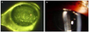
Figure 2
(Case 1) 1 week after amniotic membrane transplantation and symblepharon removal on left eye. Corneal ulcer and diffuse punctate erosion were improved.
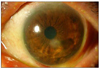
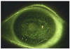
Figure 3
(Case 1) 1 month after amniotic membrane transplantation and symblepharon removal on left eye. (A) Central corneal thinning and opacity (arrow). (B) Descemetocele formation on central cornea (arrow head).

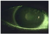
Figure 4
(Case 2) Anterior segment photograph of left eye at initial visit. (A) Filamentary keratitis and diffuse punctate corneal erosion. (B) Fine endothelial keratoprecipitates.
References
1. Wallace DJ, Hahn B. Dubois' Lupus Erythematosus. 2007. 7th ed. Philadelphia: Lippincott Williams & Wilkins;65–86.
2. Hahn BH. Fauci AS, Braunwald E, Hauser S, editors. Systemic lupus erythematosus. Harrison's Principles of Internal Medicine. 2008. v. 2:17th ed. New York: McGraw-Hill;chap. 313.
3. Tan EM, Cohen AS, Fries JF, et al. The 1982 revised criteria for the classification of systemic lupus erythematosus. Arthritis Rheum. 1982. 25:1271–1277.
4. Sivaraj RR, Durrani OM, Denniston AK, et al. Ocular manifestations of systemic lupus erythematosus. Rheumatology (Oxford). 2007. 46:1757–1762.
5. Jensen JL, Bergem HO, Gilboe IM, et al. Oral and ocular sicca symptoms and findings are prevalent in systemic lupus erythematosus. J Oral Pathol Med. 1999. 28:317–322.
6. Read RW. Clinical mini-review: systemic lupus erythematosus and the eye. Ocul Immunol Inflamm. 2004. 12:87–99.
7. Kim IT, Chang SD. Papilledema and cerebral venous thrombosis in a patient with systemic lupus erythematosis. J Korean Ophthalmol Soc. 1999. 40:2015–2019.
8. Hwang HS, Kim DH. Transient myopia with severe chemosis associated with systemic lupus erythematosus. J Korean Ophthalmol Soc. 2007. 48:1445–1448.
9. Im CY, Kim SS, Kim HK. Bilateral optic neuritis as first manifestation of systemic lupus erythematosus. Korean J Ophthalmol. 2002. 16:52–58.
10. Shin SY, Lee JM. A case of multiple cranial nerve palsies as the initial ophthalmic presentation of antiphospholipid syndrome. Korean J Ophthalmol. 2006. 20:76–78.
11. Oh PC, Kim GH, Jin CH, Baek HJ. A case of systemic lupus erythematous associated with neuromyelitis optica (Devic's Syndrome). J Korean Rheum Assoc. 2007. 14:263–267.
12. Yun KA, Kim JG. A case of orbital myositis secondary to systemic lupus erythematosus. J Korean Rheum Assoc. 2006. 13:171–176.
13. Kim IT, Na SC, Lee KJ. Vascular occlusions associated with antiphospholipid antibodies in systemic lupus erythematosus. J Korean Ophthalmol Soc. 2000. 41:427–432.
14. Park DH, Chung SK, Koo HM. A case of optic neuritis and central retinal vein occlusion associated with systemic lupus erythematosus. J Korean Ophthalmol Soc. 1994. 35:116–121.
15. Jung NH, Kim SY. A case of severe retinal vaso-occlusive disease in systemic lupus erythematosus. J Korean Ophthalmol Soc. 1993. 34:1287–1292.
16. Duke-Elder S, Leigh AG. System of Ophthalmology: Diseases of the Outer Eye. Part 2. 1965. Vol. 8. St Louis: Mosby.
17. Tanioka H, Yokoi N, Komuro A, et al. Investigation of the corneal filament in filamentary keratitis. Invest Ophthalmol Vis Sci. 2009. 50:3696–3702.
 XML Download