

 PDF
PDF ePub
ePub Citation
 Print
Print


Abstract
Purpose
To report two cases of spontaneous suprachoroidal hemorrhage associated with wet type of age-related macular degeneration (ARMD) and systemic hypertension.
Case summary
Two women, aged 76 and 73 years, with a history of systemic hypertension for more than ten years had been treated in an eye clinic for wet type of ARMD. The 76-year-old woman was scheduled to receive an injection of intravitreal Lucentis® but experienced sudden onset loss of vision and ocular pain in her left eye. The 73-year-old woman had received no specific treatment for wet type of ARMD. She also complained of visual disturbance and ocular pain. In these two cases, slit lamp or B-scan examination disclosed suprachoroidal hemorrhage. Surgical intervention relieved the ocular pain and symptoms in both cases.
Conclusions
Even if a patient has not received systemic anticoagulation/thrombolytic therapy, if wet-type ARMD patients are elderly and have systemic hypertension, they should receive regular follow-ups because old age and systemic hypertension are risk factors of spontaneous suprachoroidal hemorrhage.
References
1. Chu TG, Green RL. Suprachoroidal hemorrhage. Surv Ophthalmol. 1999; 43:471–86.

2. Pfingst AO. Expulsive choroidal hemorrhage complicating cataract surgery. South Med J. 1936; 29:323.
3. Terson A. Hemorragies sous-choroidiennes traumatiques et expulsives. Arch Ophtalmol. 1907; 27:446.
4. Verhoeff FH. Scleral puncture for expulsive subchoroidal hemorrhage following sclerotomy. Ophthalmic Res. 1915; 24:55–9.
5. Barsam A, Heatley CJ, Herbert L. Spontaneous suprachoroidal hemorrhage secondary to thrombolysis for the treatment of myocardial infarction. Clin Experiment Ophthalmol. 2006; 34:177–9.
6. Maumenee AE, Schwartz MF. Acute intraoperative choroidal effusion. Am J Ophthalmol. 1985; 100:147–54.
7. Zauberman H. Expulsive choroidal haemorrhage: an experimental study. Br J Ophthalmol. 1982; 66:43–5.
8. Beyer CF, Peyman GA, Hill JM. Expulsive choroidal hemorrhage in rabbits. A histopathologic study. Arch Ophthalmol. 1989; 107:1648–53.
9. Davison JA. Acute intraoperative suprachoroidal hemorrhage in capsular bag phacoemulsification. J Cataract Refract Surg. 1993; 19:534–7.
10. Duncker GI, Rochels R. Delayed suprachoroidal hemorrhage after penetrating keratoplasty. Int Ophthalmol. 1995–1996; 19:173–6.
11. Cantor LB, Katz LJ, Spaeth GL. Complications of surgery in glaucoma. Suprachoroidal expulsive hemorrhage in glaucoma patients undergoing intraocular surgery. Ophthalmology. 1985; 92:1266–70.
12. Fastenberg DM, Perry HD, Donnenfeld ED, et al. Expulsive suprachoroidal hemorrhage with scleral buckling surgery. Arch Ophthalmol. 1991; 109:323.
13. Koh T, Jung J-Y, Shim HS, Kim HK. Delayed suprachoroidal hemorrhage after ahmed valve implantation for neovascular glaucoma. J Korean Ophthalmol Soc. 2009; 50:635–9.
14. Lim HW, Ko BW, Song Y, Lee BR. Suprachoroidal hemorrhage during pars plana vitrectomy associated with valsalva maneuver. J Korean Ophthalmol Soc. 2008; 49:1022–7.
15. Chorich LJ, Derick RJ, Chambers RB, et al. Hemorrhagic ocular complications associated with the use of systemic thrombolytic agents. Ophthalmology. 1998; 105:428–31.
16. Chak M, Williamson TH. Spontaneous suprachoroidal haemorrhage associated with high myopia and aspirin. Eye. 2003; 17:525–7.
17. Wong JS. Spontaneous suprachoroidal haemorrhage in a patient receiving low-molecular-weight heparin (fraxiparine) therapy. Aust N Z J Ophthalmol. 1999; 27:433–4.
18. Chen YY, Chen YY, Sheu SJ. Spontaneous suprachoroidal hemorrhage associated with age-related macular degeneration and anti-coagulation therapy. J Chin Med Assoc. 2009; 72:385–7.
19. Knox FA, Johnston PB. Spontaneous suprachoroidal haemorrhage in a patient with age-related macular degeneration on excessive anticoagulation therapy. Eye. 2002; 16:669–70.
20. Chandra A, Barsam A, Hugkulstone C. A spontaneous suprachoroidal haemorrhage: a case report. Cases J. 2009; 2:185.
21. Yang SS, Fu AD, McDonald HR, et al. Massive spontaneous choroidal hemorrhage. Retina. 2003; 23:139–44.
Figure 1.
(A) Optical coherence tomography (OCT) of her left eye shows subretinal fluid and retinal pigment epithelial detachment. (B) Early phase of angiography shows dotlike hyperfluorescence at 1 o'clock and 4 o'clock positions of the central avascular zone with massive subretinal hemorrhage. (C) Late phase of angiography reveals massive leakage of fluorescence at the site with dotlike leakage in the early phase. And to conclude, there was subretinal neovascularization in the macular area and it caused massive subretinal hemorrhage.
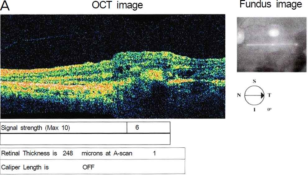
Figure 2.
(A) Axial B-mode scan. Suprachoroidal space was filled with hyperreflective and homogeneous echo structure. A-mode scan shows tall wave in front of retinal wave (Indicated with arrow on the A mode study). On both scans, these findings mean suprachoroidal hemorrhage. (B) Fundus photograph reveals kissing retina.
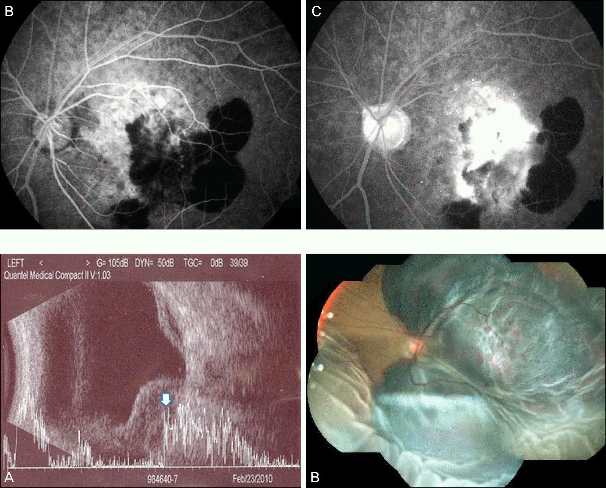
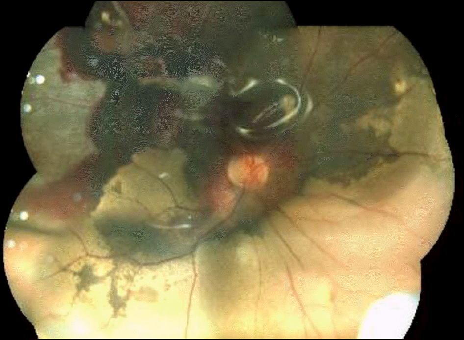
Figure 3.
After suprachoroidal hemorrhage drainage, the retina has stable condition with good silicone oil tamponade.
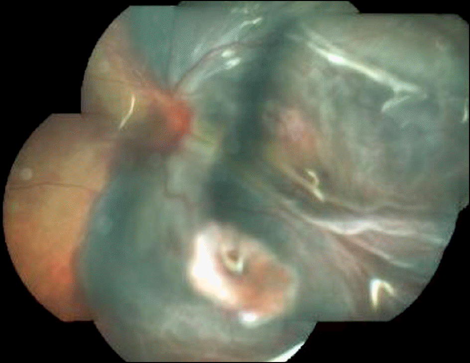
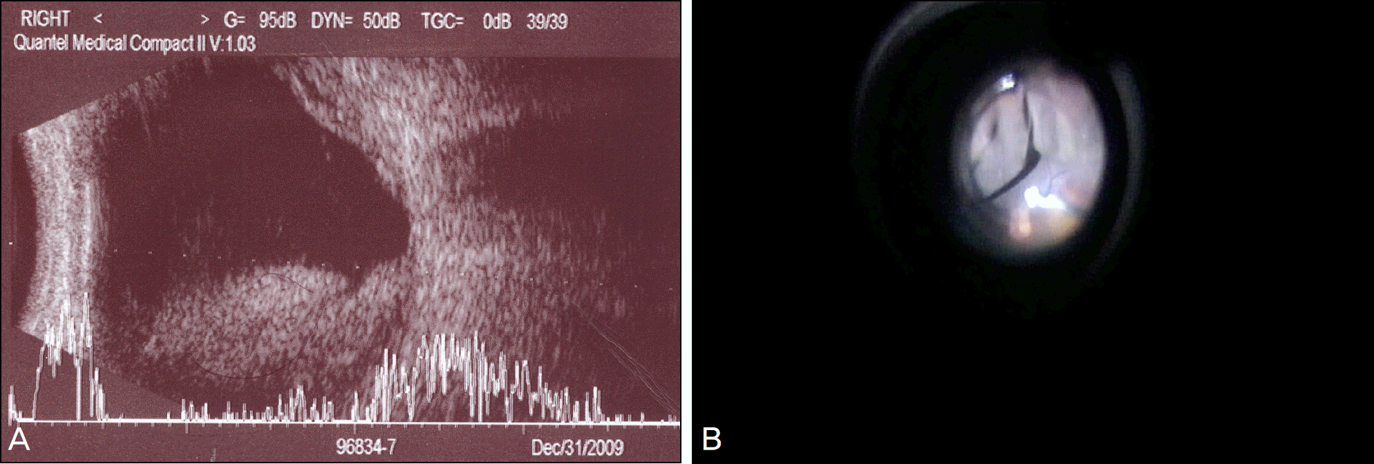
Figure 4.
(A) Axial B-mode scan. Vitreous cavity and suprachoroidal space were filled with hyperreflective and homogeneous echo structure. These findings mean vitreous hemorrhage and suprachoroidal hemorrhage. (B) After vitreous hemorrhage removal, retina shows kissing retina which means suprachoroidal hemorrhage.
 XML Download