Abstract
Purpose
To report a new modified method using a mixture of sodium hyaluronate and indocyanine green solution to facilitate the complete removal of a large conjunctival cyst.
Case summary
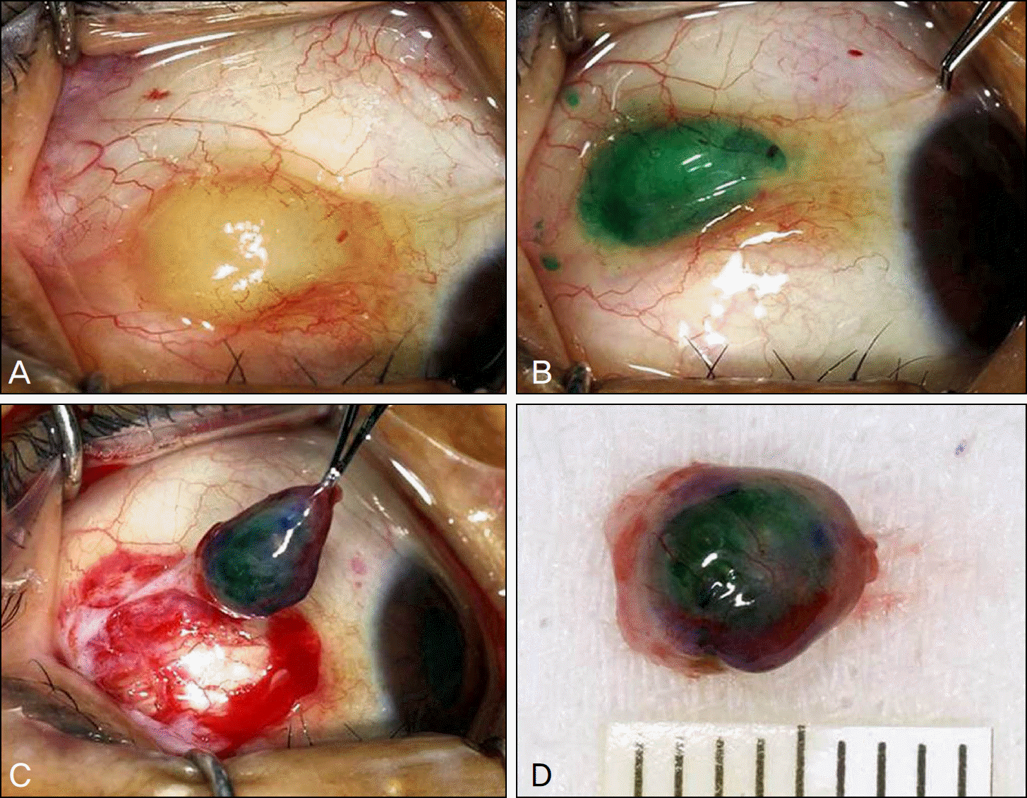
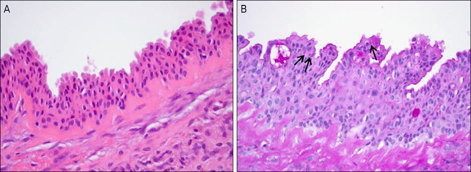
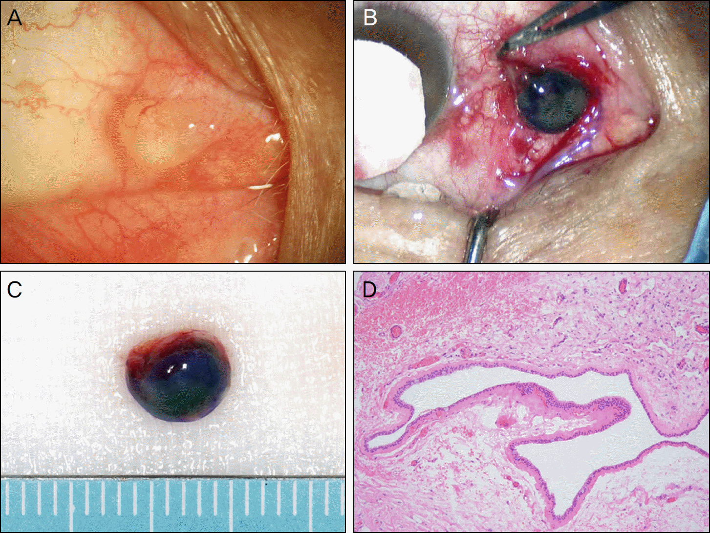
Two patients with a large conjunctival cyst on the bulbar conjunctiva were treated. In order to achieve complete removal, a mixture of 1% sodium hyaluonate and indocyanine green solution was injected through a 27-G needle into the cyst. The procedure provided excellent visualization of the cyst boundaries while maintaining cyst integrity allowing for an easy and complete resection. Apocrine hidrocystoma and a simple retention cyst were confirmed on histopathologic examination, respectively.
Go to : 
References
2. Cherrick GR, Stein SW, Leevy CM, Davidson CS. Indocyanine green: observations on its physical properties, plasma decay, and hepatic extraction. J Clin Invest. 1960; 39:592–600.

3. Kogure K, David NJ, Yamanouchi U, Choromokos E. Infrared absorption angiography of the fundus circulation. Arch Ophthalmol. 1970; 83:209–14.

4. Horiguchi M, Miyake K, Ohta I, Ito Y. Staining of the lens capsule for circular continuous capsulorrhexis in eyes with white cataract. Arch Ophthalmol. 1998; 116:535–7.

5. Pandey SK, Werner L, Escobar-Gomez M, et al. Dye-enhanced cataract surgery. Part 1: anterior capsule staining for capsulorhexis in advanced/white cataract. J Cataract Refract Surg. 2000; 26:1052–9.
6. Da Mata AP, Burk SE, Riemann CD, et al. Indocyanine green-as-sisted peeling of the retinal internal limiting membrane during vitrectomy surgery for macular hole repair. Ophthalmology. 2001; 108:1187–92.

7. Li K, Wong D, Hiscott P, et al. Trypan blue staining of internal limiting membrane and epiretinal membrane during vitrectomy: visual results and histopathological findings. Br J Ophthalmol. 2003; 87:216–9.

8. Hahm IR, Tae KS, Cho SW, et al. The outcomes after indocyanine green-assisted peeling of the internal limiting membrane in macular hole surgery. J Korean Ophthalmol Soc. 2005; 46:1361–7.
9. Auffarth GU, Holzer MP, Vissesook N, et al. Removal times and techniques of a viscoadaptive ophthalmic viscosurgical device. J Cataract Refract Surg. 2004; 30:879–83.

10. Kobayashi A, Saeki A, Nishimura A, et al. Visualization of conjunctival cyst by indocyanine green. Am J Ophthalmol. 2002; 133:827–8.

11. Hoffman RS, Fine IH, Packer M. Stabilization of flat anterior chamber after trabeculectomy with Healon5. J Cataract Refract Surg. 2002; 28:712–4.

12. Kobayashi A, Sugiyama K. Visualization of conjunctival cyst using Healon V and trypan blue. Cornea. 2005; 24:759–60.

Go to : 
 | Figure 1.(A) 6×5 mm-sized large conjunctival cyst is seen in the bulbar conjunctiva of the right eye.(B) The cyst margin are clearly visible through the conjunctiva after stained with a mixture of sodium hyaluronate and indocyanine green solution. (C, D) Successful removal of the conjunctival cyst with delineated capsule and preserved integrity. |
 | Figure 2.Double layered lining of apocrine hidrocytoma (H&E stain, ×40) (A), consisting of large columnar cells with eosinophilic cytoplasm with luminal decapitation secretion in the inner layer and flat myoepithelial cells in the outer layer (H&E stain, ×100).(B) Arrows indicate PAS-positive, diastase-resistant granule on the apical surfaces of the inner layer (PAS stain, ×100). |
 | Figure 3.(A) 6×4 mm-sized large conjunctival cyst is seen in the medial conjunctiva adjacent to the caruncle of the right eye. (B) The cyst is stained using a mixture of sodium hyaluronate and indocyanine green solution. (C) Succesful removal of the conjunctival cyst with delineated capsule and preserved integrity. (D) A solitary uniocular cyst is lined by non-keratinizing cuboidal epithelium. Goblet cells are often included (H&E stain, ×20). |


 PDF
PDF ePub
ePub Citation
 Print
Print


 XML Download