

 PDF
PDF ePub
ePub Citation
 Print
Print


Abstract
Purpose
To report a case of endophthalmitis treated with surgical removal of the inflammatory endothelial plaque.
Case summary
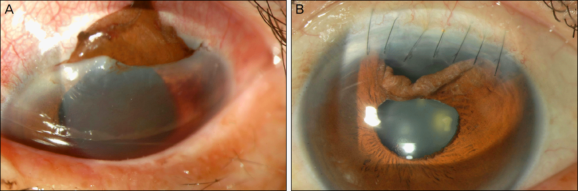
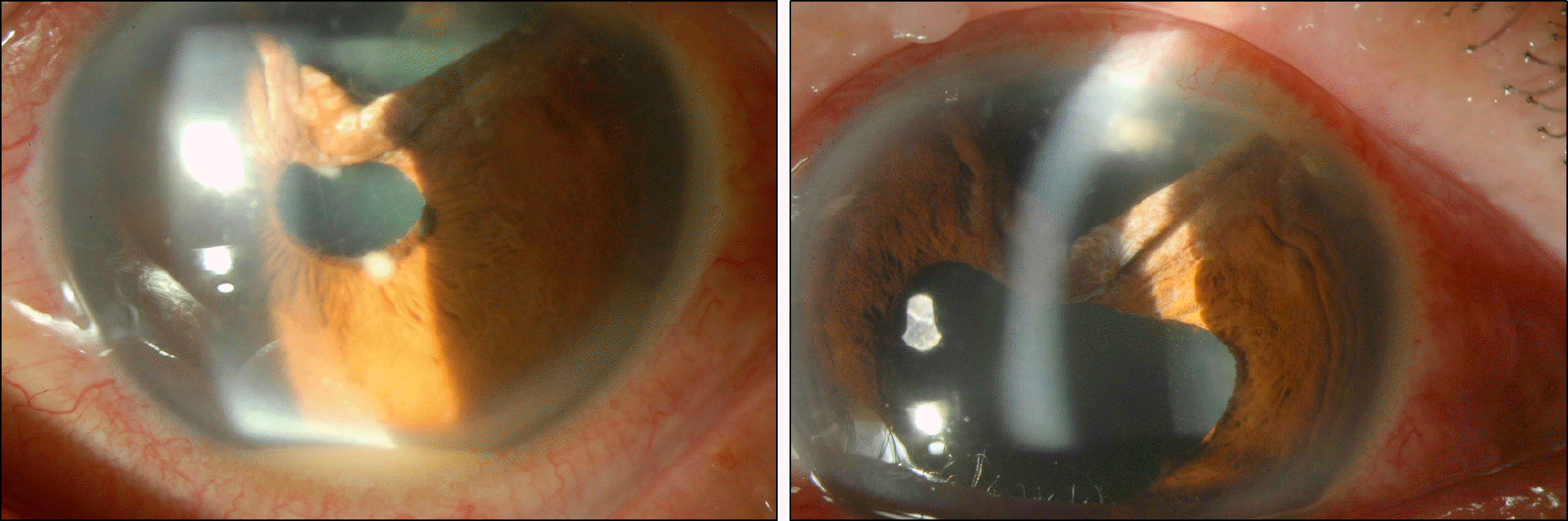
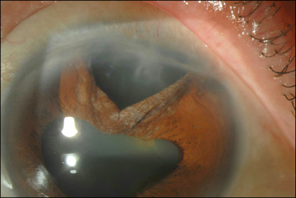
A 61-year-old male was transferred to our clinic due to corneal laceration of the left eye. An emergency operation for the lacerated cornea was performed. After the operation, the patient had no specific symptoms for 8 months but then visited our clinic with sudden decreased visual acuity. On slit lamp examination, the patient had some chamber reactions. Anterior chamber reactions exacerbated after 2 months and the best corrected visual acuity was decreased from 1.0 to 0.08. An inflammatory corneal endothelial plaque and endothelial precipitates had developed. The posterior segment was not visualized due to the severe anterior chamber inflammatory reaction. No growth was observed on bacterial or fungal cultures. However, administration of eye drops and oral voriconazole were initiated based on a clinical impression suspicious of fungal infection. Despite the treatment, the infection did not respond. Voriconazole was then directly injected into the vitreous and anterior chamber. Although the patient's best corrected visual acuity slightly improved, the inflammatory reactions of the anterior chamber and vitreous did not. The inflammatory endothelial plaque on the patient's cornea was then surgically removed and the best corrected visual acuity improved to 1.0. Mycelium was detected on the KOH smear of the endothelial plaque. There were no further inflammatory reactions in the anterior chamber or vitreous after surgical removal of the endothelial plaque.
References
1. Pettit TH, Olson RJ, Foos RY, Martin WJ. Fungal endophthalmitis following intraocular lens implantation. A surgical epidemic. Arch Ophthalmol. 1980; 98:1025–39.
2. Lee SJ, Lee JJ, Kim SD. Topical and oral voriconazole in the treatment of fungal keratitis. Korean J Ophthalmol. 2009; 23:46–8.

3. Kramer M, Kramer MR, Blau H, et al. Intravitreal voriconazole for the treatment of endogenous Aspergillus endophthalmitis. Ophthalmology. 2006; 113:1184–6.
4. Srinivasan M, Gonzales CA, George C, et al. Epidemiology and aetiological diagnosis of corneal ulceration in Madurai, south India. Br J Ophthalmol. 1997; 81:965–71.
5. Wykoff CC, Flynn HW Jr, Miller D, et al. Exogenous fungal endophthalmitis: microbiology and clinical outcomes. Ophthalmology. 2008; 115:1501–7.
6. Axelrod AJ, Peyman GA, Apple DJ. Toxicity of intravitreal injection of amphotericin B. Am J Ophthalmol. 1973; 76:578–83.
7. Alexandridou A, Reginald AY, Stavrou P, Kirkby GR. Candida endophthalmitis after tattooing in an asplenic patient. Arch Ophthalmol. 2002; 120:518–9.
8. Biju R, Sushil D, Georgy NK. Successful management of presumed Candida endogenous endophthalmitis with oral voriconazole. Indian J Ophthalmol. 2009; 57:306–8.
9. Kim KH, Kim MJ, Tchah HW. Management of fungal ocular infection with topical and intracameral voriconazole. J Korean Ophthalmol Soc. 2008; 49:1054–60.
10. Yoon JU, Kim SW, Ha BJ, et al. A case of fungal keratitis treated with voriconazole. J Korean Ophthalmol Soc. 2008; 49:1680–4.
11. Oh DH, Kim JC, Chun YS. A case of fusarium deep keratitis following scleral graft. J Korean Ophthalmol Soc. 2010; 51:606–10.
 XML Download