Abstract
Purpose
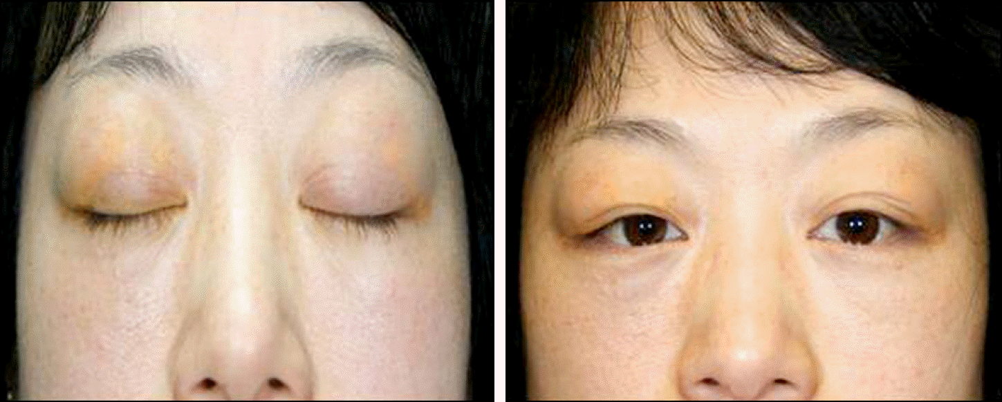
To report a case of adult orbital xanthogranulomatous disease presented as bilateral swelling and yellowish eyelids in a 42-year-old woman who was misdiagnosed with xanthelasma at a dermatologic clinic.
Case summary
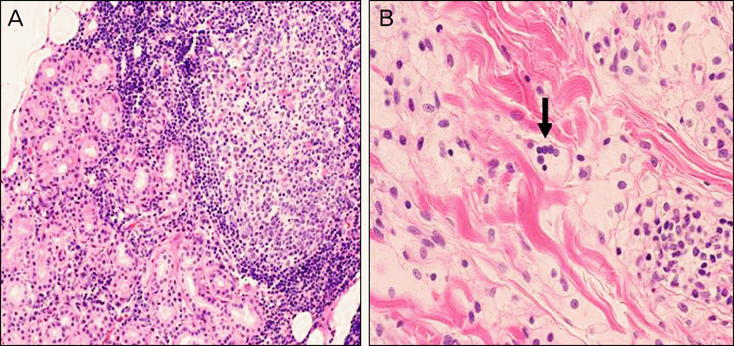
A 42-year-old woman presented yellowish and swollen eyelids in both eyes of 2.5 year duration. She had no past history of systemic diseases or other ophthalmologic problems. MRI showed heterogeneous eyelid masses and hypertrophic changes of the lacrimal glands in both eyes. There were no abnormalities on chest X-ray exams. The blood cholesterol and low density lipoprotein levels were increased. Incisional biopsy showed many foamy histiocytes, lymphocytes with germinal centers, several touton giant cells, and negative S100/CD 1 staining; all being features consistent with adult onset xanthogranulomatous disease.
Go to : 
References
1. Sivak-Callcott JA, Rootman J, Rasmussen SL, et al. Adult xanthogranulomatous disease of the orbit and ocular adnexa: new immunohistochemical findings and clinical review. Br J Ophthalmol. 2006; 90:602–8.

2. Guo J, Wang J. Adult orbital xanthogranulomatous disease: review of the literature. Arch Pathol Lab Med. 2009; 133:1994–7.

3. Choi HJ, Kwon HJ, Kim HY. A case of posttraumatic necrobiotic xanthogranuloma of the orbit. J Korean Ophthalmol Soc. 2008; 49:677–80.

4. Park SH, Rah SH, Kim YH. Juvenile xanthogranuloma as an isolated corneoscleral limbal mass: a case report. Korean J Ophthalmol. 2003; 17:63–6.

5. Lee KM, Kim NJ. Juvenile xanthogranuloma presenting as a nodule of the eyelid. J Korean Ophthalmol Soc. 2008; 49:1173–6.

6. Frankel RM, Capone R. Xanthelasmas and xanthomas ― cutaneous clues to systemic lipid disorders. Clin Eye Vis Care. 1995; 7:117–28.

7. Hammond MD, Niemi EW, Ward TP, Eiseman AS. Adult orbital xanthogranuloma with associated adult-onset asthma. Ophthal Plast Reconstr Surg. 2004; 20:329–32.

Go to : 
 | Figure 1.Clinical photographs of the patient's eyelid before starting treatment. These pictures show bilateral yellowish eyelid swelling. |
 | Figure 2.MRI demonstrated bilateral soft tissue swelling with no bone invasions. (A) T1 image show a 5-mm ovoid hypointense lesion (arrow) at the left upper eyelid, midline. (B) T2 axial image show diffuse swelling with heterogenous signal intensities at bilateral lacrimal glands. (C) T1 axial image show heterogenous soft tissue swelling at bilateral eyelids and preseptal areas. |
 | Figure 3.Histologic findings of orbital mass. (A) Variable germinal centers and nodular lymphoid infiltrates in the lacrimal gland (H&E stain, ×100). (B) Touton giant cells (arrow) and many foamy histiocytes in the subcutaneous layer (H&E stain, ×200). |
 | Figure 4.Clinical photographs of the patient's eyelids. (A) After excisional biopsy at month 4 and steroid therapy for 1.5 months. (B): After excisional biopsy at month 7 and steroid therapy for 4.5 months. These pictures show decreased yellow colored eyelids and reduction of eyelid swelling. |


 PDF
PDF ePub
ePub Citation
 Print
Print


 XML Download