Abstract
Purpose
To report a case of a patient with infectious endophthalmitis associated with necrotizing scleritis that was treated with pars plana vitrectomy and permanent amniotic membrane transplantation.
Case summary
A 76-year-old man with pain and visual loss in the right eye was diagnosed with infectious endophthalmitis and necrotizing scleritis. The visual acuity in the right eye was hand motion, and the slit lamp examination showed infection of the conjunctiva and sclera, corneal edema, hypopyon, and necrosis of nasal sclera. An intravitreal antibiotic injection was given, and Pseudomonas aeruginosa was cultured in vitreous fluid. Two days afterward, when vitrectomy was performed, leakage from the scleral microperforation and necrosis of the peripheral cornea was observed. Thus, a 10-layered permanent amniotic membrane transplantation was performed. The patient was given topical antibiotics and steroids, oral prednisolone, and cyclophosphamide postoperatively. After 74 days, endophthalmitis was remitted, and scleritis was well controlled. His visual acuity recovered to 20/40.
References
1. Jabs DA, Mudun A, Dunn JP, Marsh MJ. Episcleritis and scleritis: clinical features and treatment results. Am J Ophthalmol. 2000; 130:469–76.

3. Huang FC, Huang SP, Tseng SH. Management of infectious scleritis after pterygium excision. Cornea. 2000; 19:34–9.

4. Reynolds MG, Alfonso E. Treatment of infectious scleritis and keratoscleritis. Am J Ophthalmol. 1991; 112:543–7.

5. Kachmaryk M, Bouchard CS, Duffner LA. Bilateral fascia lata patch grafts in a patient with progressive scleromalacia perforans. Ophthalmic Surg Lasers. 1996; 27:397–400.

6. Lee SH, Tseng SC. Amniotic membrane transplantation for persistent epithelial defects with ulceration. Am J Ophthalmol. 1997; 123:303–12.

7. Tseng SC, Prabhasawat P, Lee SH. Amniotic membrane transplantation for conjunctival surface reconstruction. Am J Ophthalmol. 1997; 124:765–74.

8. Shimmura S, Shimazaki J, Ohashi Y, Tsubota K. Antiinflammatory effects of amniotic membrane transplantation in ocular surface disorders. Cornea. 2001; 20:408–13.

9. Jeoung JW, Yoon YM, Lee JL, et al. The effect of amniotic membrane transplantation on the treatment of necrotizing scleritis after pterygium excision. J Korean Ophthalmol Soc. 2004; 45:1981–8.
10. Kim YK, Kim TY. 4 Cases of pseudomonas scleritis after pterygium excision. J Korean Ophthalmol Soc. 1999; 40:2304–12.
11. Han YK, Wee WR. Use of immunosuppressant in the treatment of surgically induced necrotizing scleritis (sins) after pterygium excision. J Korean Ophthalmol Soc. 2003; 44:272–7.
12. Park SW, Lee MH, Lee JE, Lee JS. A case of methicillin resistant staphylococcus aureus scleritis after pterygium excision. J Korean Ophthalmol Soc. 2007; 48:157–61.
13. Moreno Honrado M, del Campo Z, Buil JA. A case of necrotizing scleritis resulting from pseudomonas aeruginosa. Cornea. 2009; 28:1065–6.
14. Hanada K, Shimazaki J, Shimmura S, Tsubota K. Multilayered amniotic membrane transplantation for severe ulceration of the cornea and sclera. Am J Ophthalmol. 2001; 131:324–31.

15. Nubile M, Carpineto P, Lanzini M, et al. Multilayer amniotic membrane transplantation for bacterial keratitis with corneal perforation after hyperopic photorefractive keratectomy: case report and literature review. J Cataract Refract Surg. 2007; 33:1636–40.
16. Kim HK, Park HS. Fibrin glue-assisted augmented amniotic membrane transplantation for the treatment of large noninfectious corneal perforations. Cornea. 2009; 28:170–6.

17. Results of the Endophthalmitis Vitrectomy Study. A randomized trial of immediate vitrectomy and of intravenous antibiotics for the treatment of postoperative bacterial endophthalmitis. Endophthalmitis Vitrectomy Study Group. Arch Ophthalmol. 1995; 113:1479–96.
18. Doft BH, Kelsey SF, Wisniewski SR. Additional procedures after the initial vitrectomy or tap-biopsy in the Endophthalmitis Vitrectomy Study. Ophthalmology. 1998; 105:707–16.

19. Kuhn F, Gini G. Ten years after. are findings of the Endophthalmitis Vitrectomy Study still relevant today? Graefes Arch Clin Exp Ophthalmol. 2005; 243:1197–9.
21. Feiz V, Redline DE. Infectious scleritis after pars plana vitrectomy because of methicillin-resistant Staphylococcus aureus resistant to fourth-generation fluoroquinolones. Cornea. 2007; 26:238–40.
22. Rich RM, Smiddy WE, Davis JL. Infectious scleritis after retinal surgery. Am J Ophthalmol. 2008; 145:695–9.

23. Morley AM, Pavesio C. Surgically induced necrotising scleritis following three-port pars plana vitrectomy without scleral buckling: a series of three cases. Eye (Lond). 2008; 22:162–4.

24. Wilhelmus KR. Indecision about corticosteroids for bacterial keratitis: an evidence-based update. Ophthalmology. 2002; 109:835–42.
Figure 1.
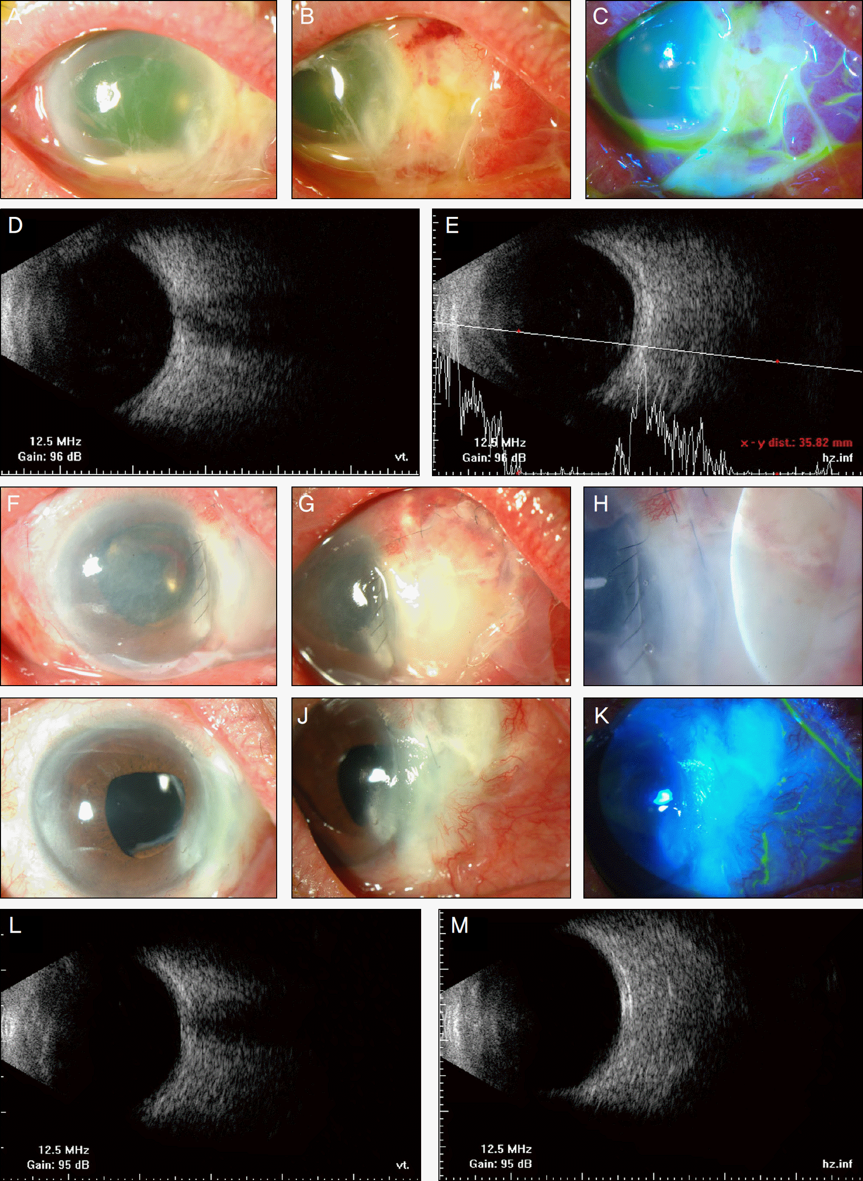
(A), (B), (C) The right eye at the first visit presented conjunctival injection, corneal edema, hypopyon, scleral edema, necrosis and thinning in nasal sclera, and epithelial defect in the nasal peripheral cornea. (D), (E) Ultrasonogram (B-scan) shows haziness in the vitreous and posterior sclera thickening at the first visit. (F), (G), (H) Amniotic membrane is intact in the necrotic nasal sclera 2 days after permanent amniotic membrane transplantation. White temporary amniotic membrane covered entire cornea and nasal conjunctiva. (I), (J), (K) At 74 days after treatment, conjunctival and scleral injection was nearly disappeared, and there is no epithelial defect in cornea and conjunctiva. (L), (M) Ultrasonogram (B-scan) shows a clear vitreous cavity and no posterior sclera thickening.


 PDF
PDF ePub
ePub Citation
 Print
Print


 XML Download